📖 Lesson: Heart Anatomy
Discover the incredible structure that keeps you alive
🎮 Interactive Games
Test your knowledge about the human heart
❤️ Structure of the Human Heart: Chambers, Valves, Vessels and Blood Flow
What the heart is and where it sits
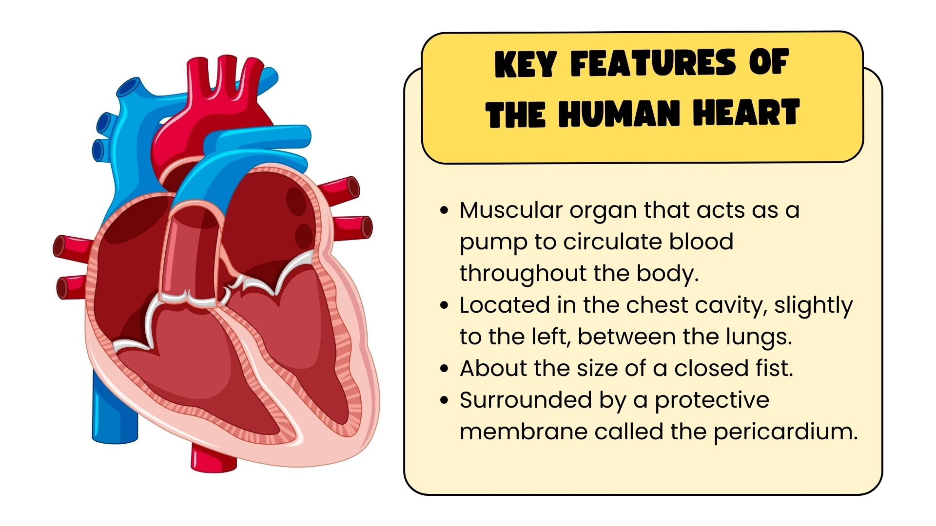
The human heart is a muscular pump about the size of your clenched fist. It lies in the chest between the two lungs, a little to the left of the midline, and is protected by the ribcage and the breastbone. Its wall is made of cardiac muscle, a special tissue that contracts rhythmically on its own and never tires, and the whole organ is wrapped in a tough double membrane called the pericardium. Its job is simple to state: it pumps blood around the body so that every cell receives oxygen and glucose and has its carbon dioxide and other wastes carried away.
One point of confusion to settle first: left and right refer to the owner's left and right, not yours. In a diagram drawn from the front, the right side of the heart appears on the left of the page.
The four chambers
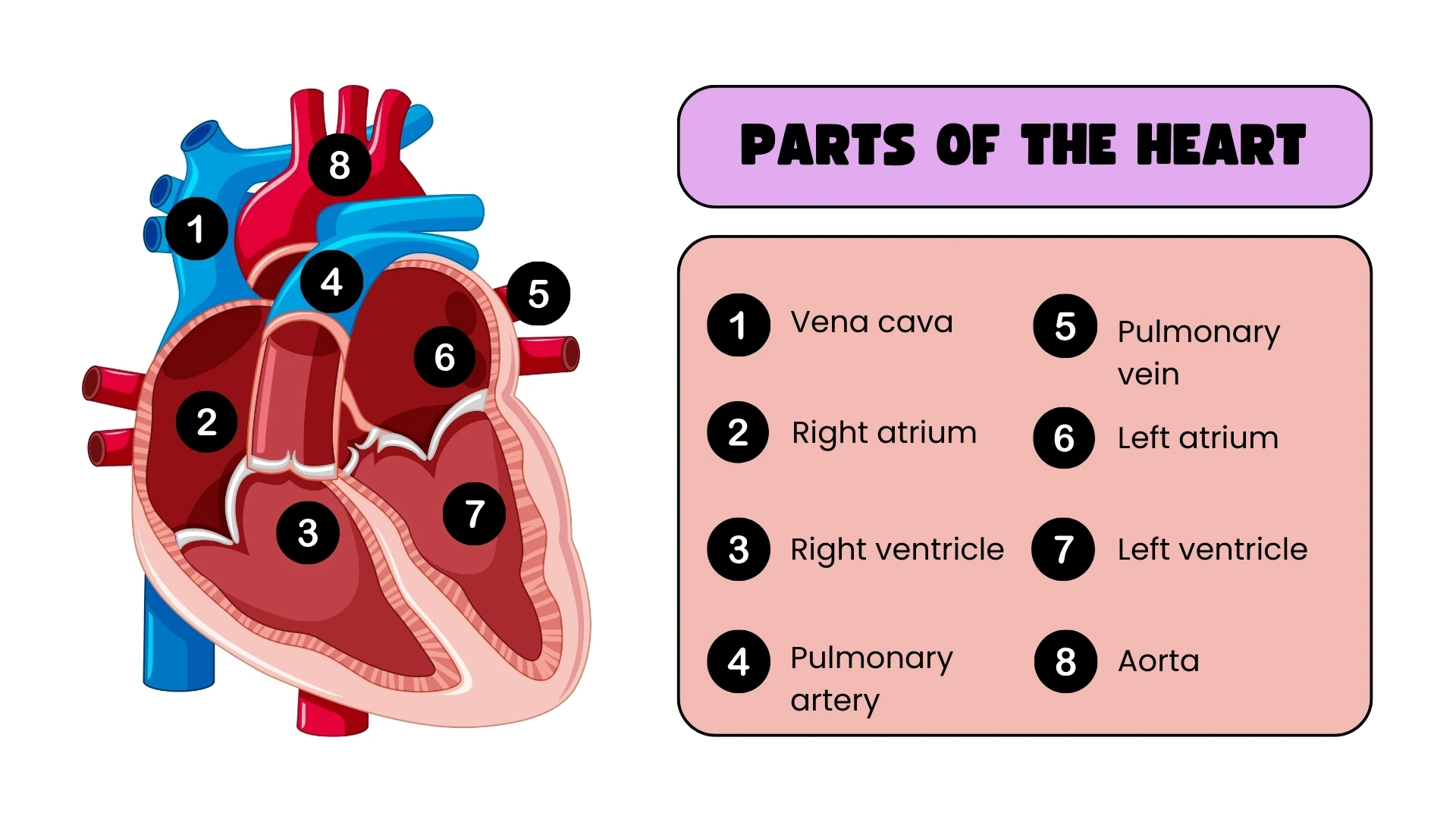
Inside, the heart is divided into four chambers. The two upper chambers are the atria (one is an atrium); the two lower chambers are the ventricles. A solid wall of muscle called the septum runs down the middle and separates the right side from the left, so blood on the two sides never mixes.
- The right atrium receives deoxygenated blood returning from the body.
- The right ventricle pumps that deoxygenated blood to the lungs.
- The left atrium receives oxygenated blood returning from the lungs.
- The left ventricle pumps oxygenated blood out to the whole body.
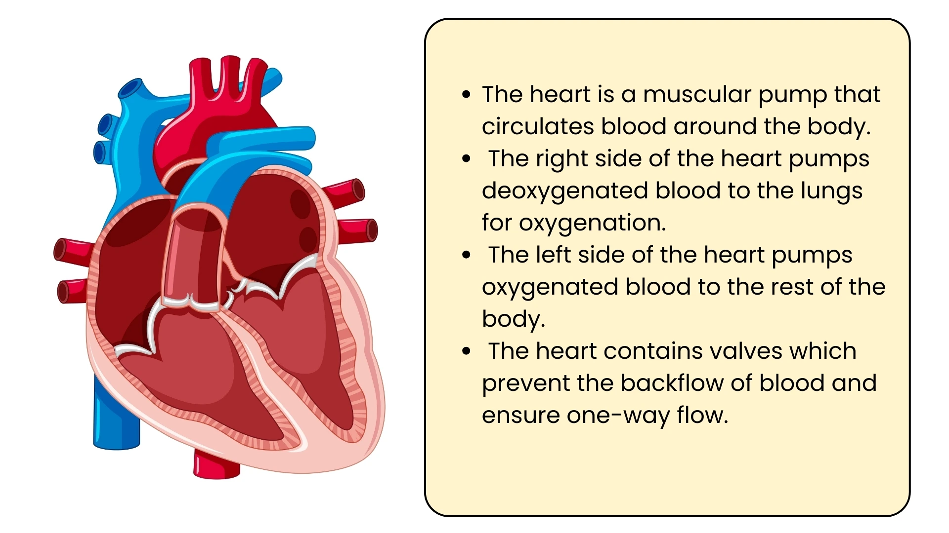
So the right side of the heart handles deoxygenated blood and the left side handles oxygenated blood. The atria have thin walls because they only push blood a short distance down into the ventricles. The ventricles have thick walls because they push blood out of the heart altogether.
Why the left ventricle has the thickest wall
Of the four chambers, the left ventricle has by far the thickest, most muscular wall. The right ventricle only has to send blood to the lungs, which lie right next to the heart, and it must do so at fairly low pressure so that the delicate capillaries in the lungs are not damaged. The left ventricle has to drive blood through the aorta to every part of the body, including the brain above it and the toes far below, and that needs a much higher pressure. A thicker wall means more muscle, and more muscle means a stronger contraction. This is a classic example of structure matching function.
The four valves
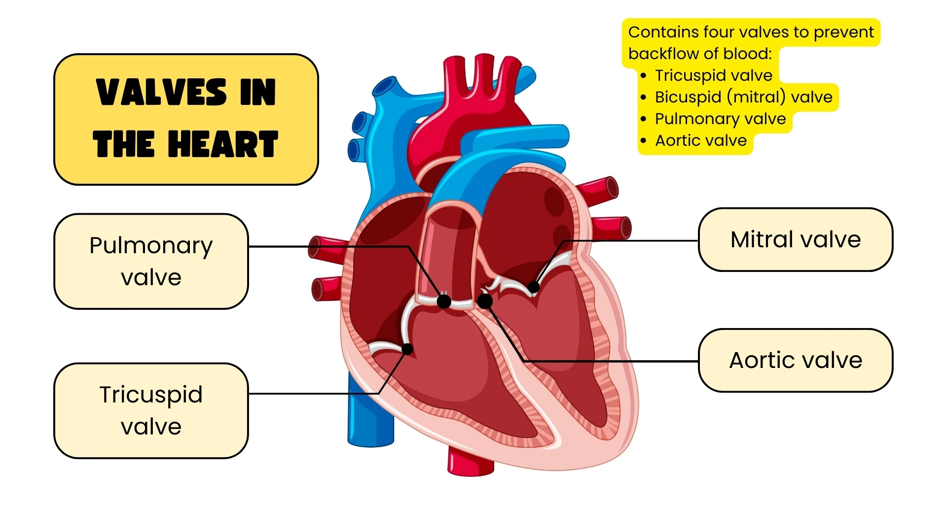
Blood must flow through the heart in one direction only, and valves make sure it does. A valve is a flap of tissue that opens when blood pushes on it from the correct side and snaps shut if blood tries to flow backwards. The heart has four:
- The tricuspid valve, between the right atrium and right ventricle, has three flaps.
- The bicuspid valve (also called the mitral valve), between the left atrium and left ventricle, has two flaps.
- The pulmonary valve sits where the right ventricle opens into the pulmonary artery.
- The aortic valve sits where the left ventricle opens into the aorta.
The tricuspid and bicuspid valves are the atrioventricular valves. They are anchored to the ventricle walls by tough cords, the tendons or chordae tendineae, which stop the flaps from being blown inside out into the atria when the ventricles contract hard. The pulmonary and aortic valves are semilunar valves, and they stop blood in the two great arteries falling back into the ventricles when the ventricles relax.
The major blood vessels
| Vessel | Type | Connects | Blood carried |
|---|---|---|---|
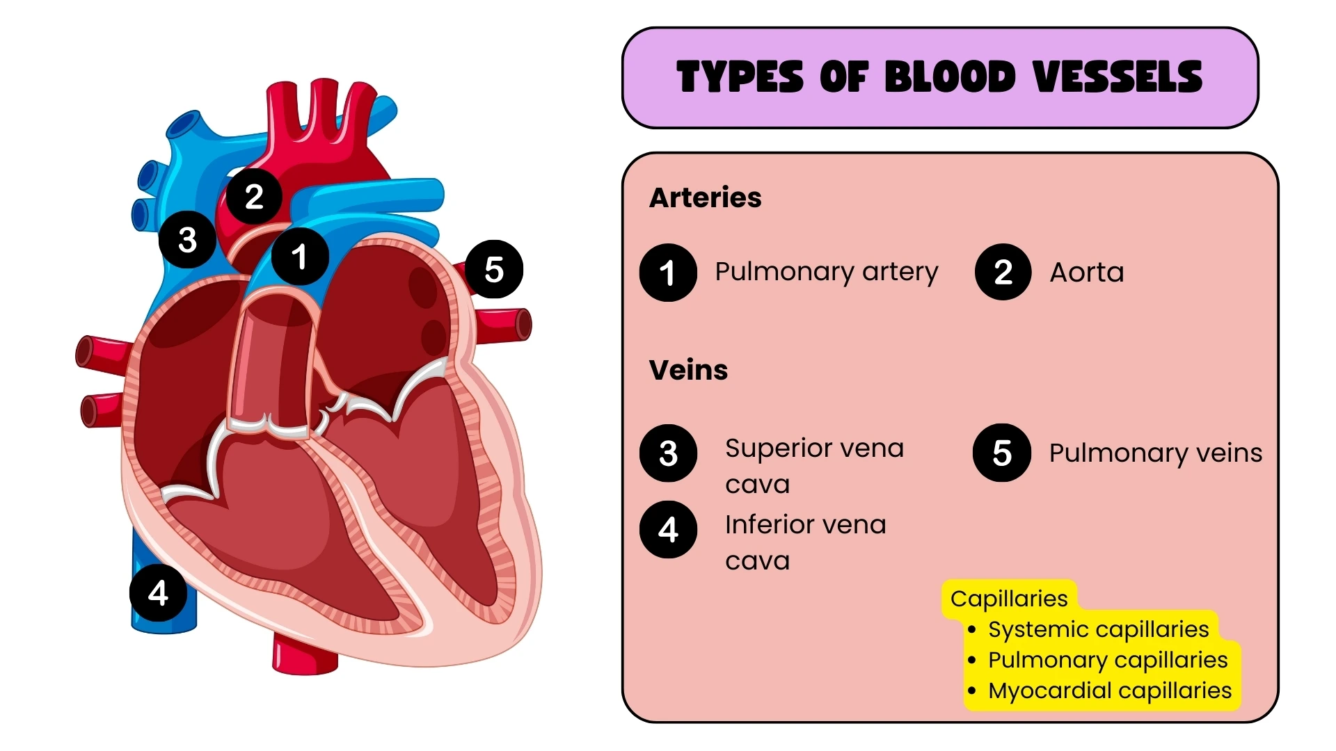
| Vena cava | Vein | Body to right atrium | Deoxygenated |
| Pulmonary artery | Artery | Right ventricle to lungs | Deoxygenated |
| Pulmonary vein | Vein | Lungs to left atrium | Oxygenated |
| Aorta | Artery | Left ventricle to body | Oxygenated |
Notice the exception in the table. Arteries normally carry oxygenated blood and veins deoxygenated blood, but the vessels between the heart and the lungs are the other way round. The pulmonary artery is the only artery in the body that carries deoxygenated blood, and the pulmonary veins are the only veins that carry oxygenated blood. An artery is defined by direction, as a vessel that carries blood away from the heart, not by the oxygen its blood contains. The heart muscle itself is supplied by its own small coronary arteries, which branch off the aorta.
How blood flows through the heart, step by step
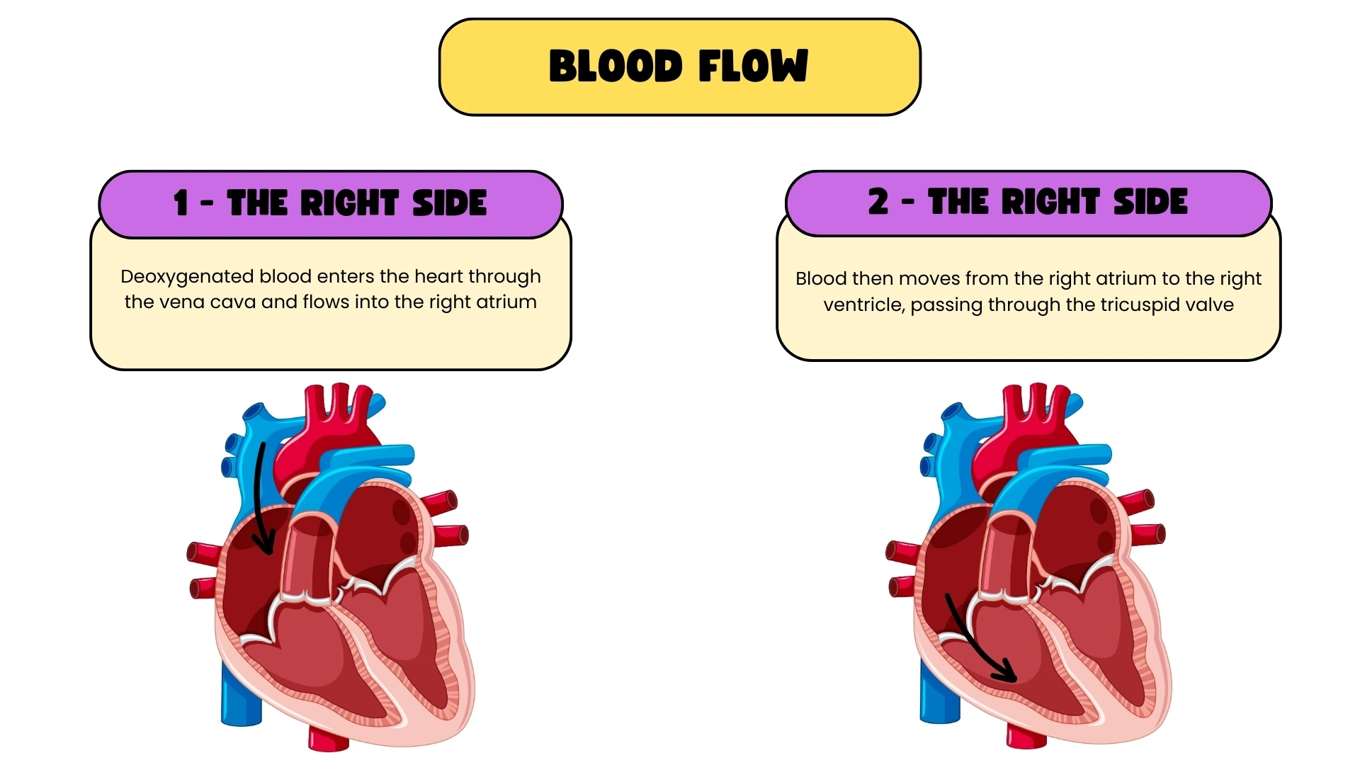
- Deoxygenated blood from the body enters the right atrium through the vena cava.
- The right atrium contracts and pushes blood through the tricuspid valve into the right ventricle.
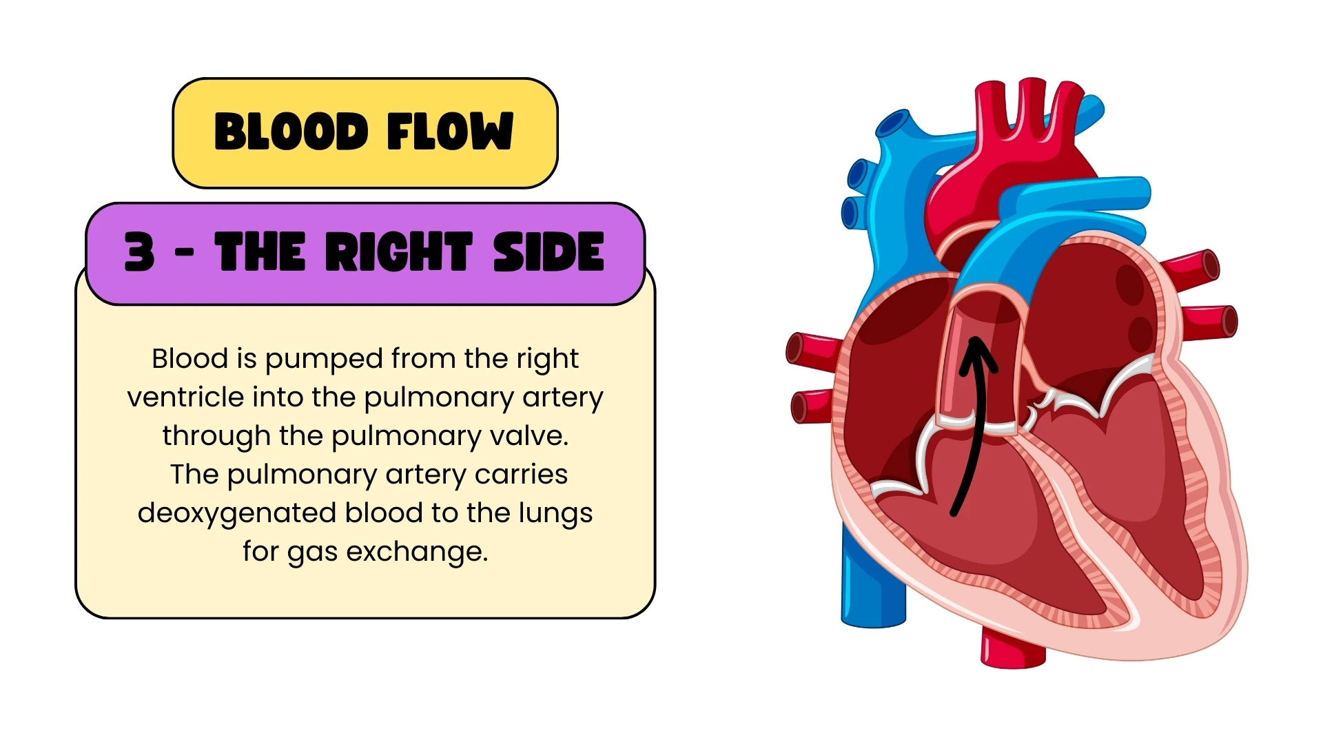
- The right ventricle contracts and pumps blood through the pulmonary valve into the pulmonary artery, which carries it to the lungs.
- In the lungs the blood picks up oxygen and gives up carbon dioxide.
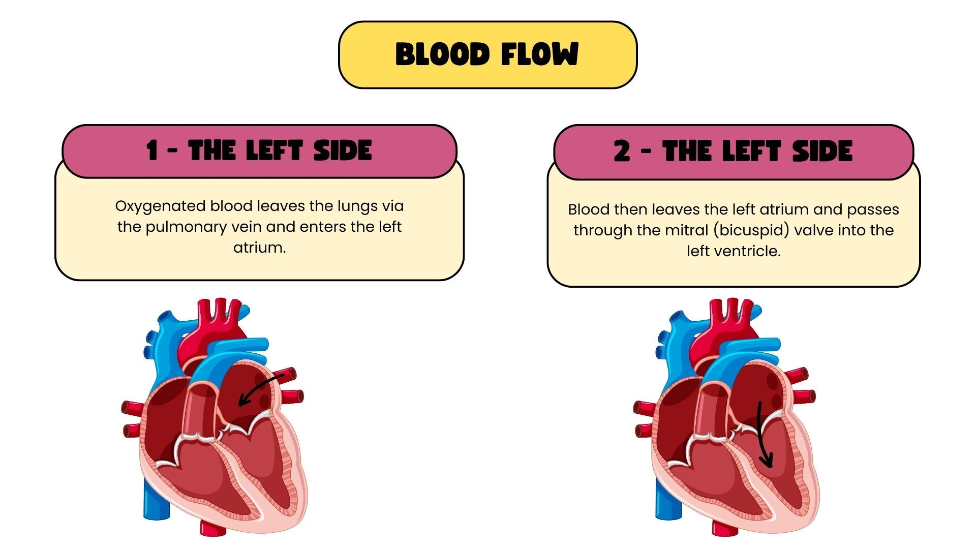
- Oxygenated blood returns through the pulmonary veins to the left atrium.
- The left atrium contracts and pushes blood through the bicuspid valve into the left ventricle.
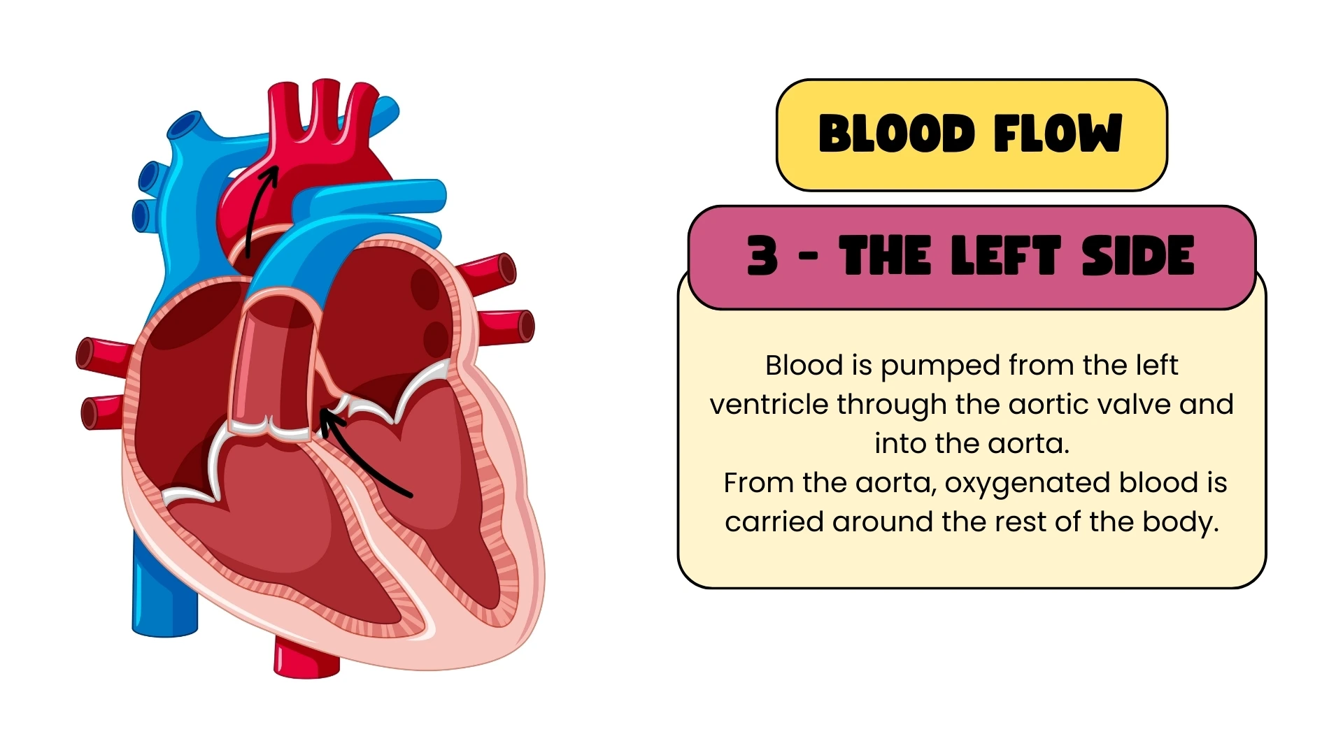
- The left ventricle contracts powerfully and pumps blood through the aortic valve into the aorta, which distributes it to the whole body.
The two atria contract together, then the two ventricles contract together, so both sides of the heart work at the same time.
Double circulation
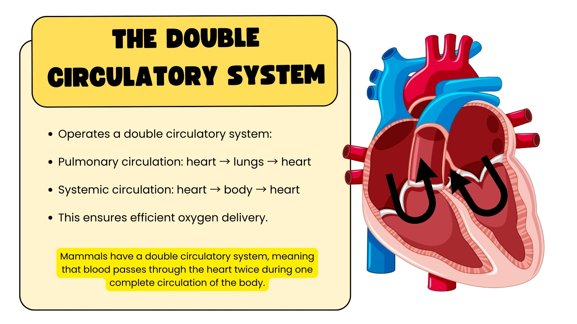
Humans have a double circulatory system: in one complete trip around the body, blood passes through the heart twice. The pulmonary circulation takes blood from the right side of the heart to the lungs and back to the left side. The systemic circulation takes blood from the left side of the heart to the rest of the body and back to the right side. The advantage of the double system is pressure: blood loses pressure as it squeezes through the lung capillaries, and returning it to the heart lets the left ventricle give it a fresh, powerful push before it sets off to the body. That keeps oxygenated blood moving quickly, which suits an active, warm-blooded animal.
Key terms
- Atrium
- One of the two thin-walled upper chambers of the heart that receive blood from the veins.
- Ventricle
- One of the two thick-walled lower chambers that pump blood out of the heart.
- Septum
- The muscular wall that separates the right and left sides of the heart.
- Valve
- A flap of tissue that allows blood to flow in one direction only.
- Vena cava
- The large vein that returns deoxygenated blood from the body to the right atrium.
- Aorta
- The largest artery, carrying oxygenated blood from the left ventricle to the body.
- Pulmonary
- Relating to the lungs; the pulmonary artery and veins run between the heart and the lungs.
- Double circulation
- A system in which blood passes through the heart twice on each complete circuit of the body.