The airways, the muscles of breathing, alveoli adaptations and gas exchange, explained step by step for exams.
Introduction: The One Thing You Cannot Postpone
You can survive several weeks without food. You can survive a few days without water. You can survive only a few minutes without oxygen. That single fact tells you how important the respiratory system is.
The remarkable thing is that you do this vital job about twenty thousand times a day without noticing it. Right now, as you read this sentence, you are breathing, and you were not thinking about it until I mentioned it. Your body handles it automatically, adjusting the speed and depth to match whether you are sleeping, walking or sprinting for a bus.
This guide explains how the respiratory system works, from the moment air enters your nose to the moment oxygen slips into your blood, and then why breathing out is just as important as breathing in.
What Is the Respiratory System?
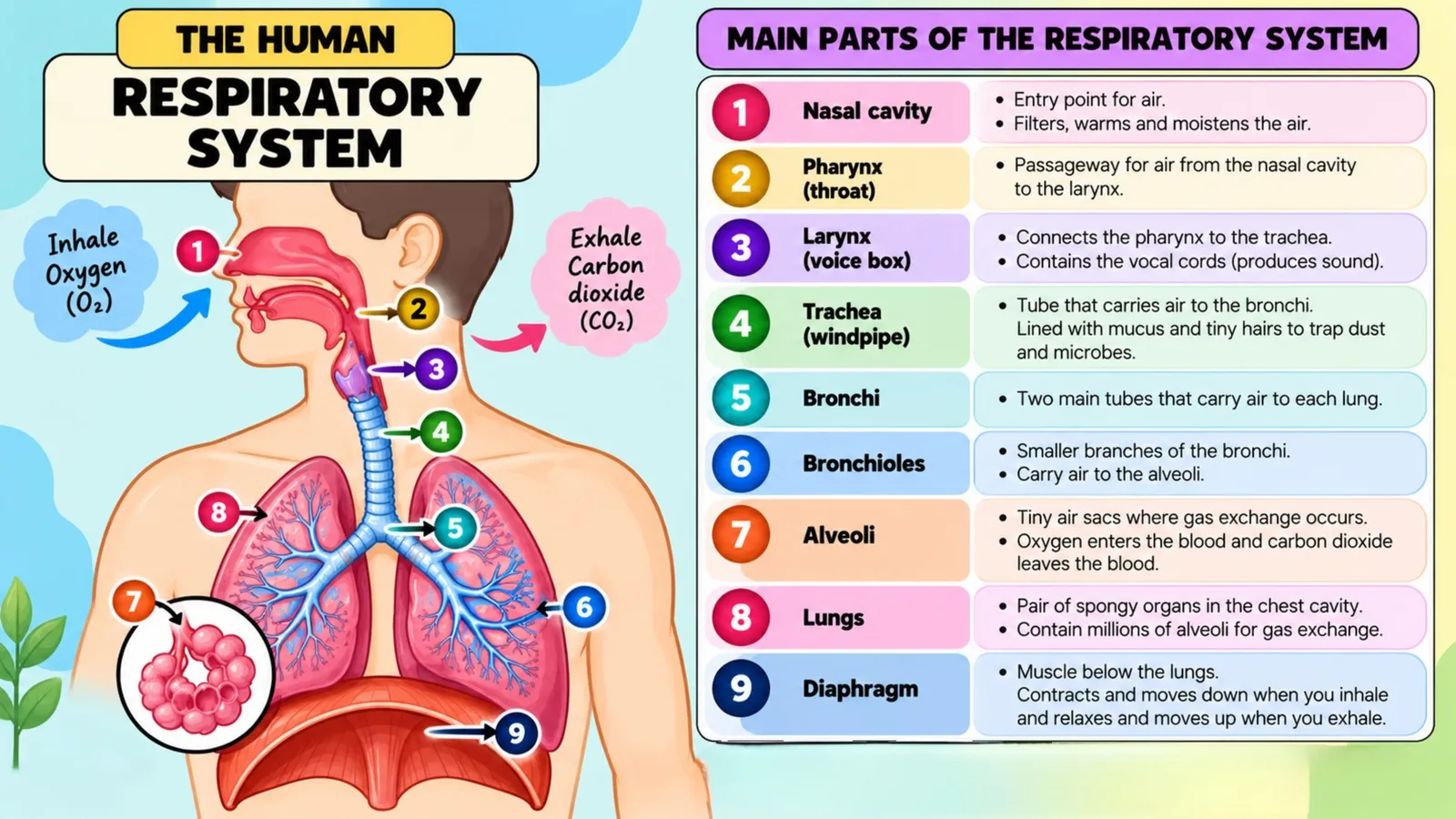
The respiratory system is the group of organs that brings oxygen into the body and removes carbon dioxide from it. It includes the nose, nasal cavity, pharynx, larynx, trachea, bronchi, bronchioles, alveoli and lungs, supported by the diaphragm and the rib muscles.
Before going further, one distinction must be made very clearly, because examiners test it constantly.
Breathing is the physical movement of air into and out of the lungs. It is a mechanical process involving muscles, and it happens in the chest.
Respiration is the chemical process inside cells where glucose is broken down to release energy. It happens in the mitochondria of every living cell, and it never stops.
Breathing supplies the oxygen that cellular respiration needs and carries away the carbon dioxide it produces. So breathing serves respiration, but the two words are not interchangeable. A person on a ventilator is being helped to breathe, but their cells are still respiring on their own.
🫁
Slides: The human respiratory system
The organs that take in oxygen and remove carbon dioxide
1/2
Why the Body Needs Oxygen
Your cells run on energy, and that energy comes from breaking down glucose. The equation for aerobic respiration is simple to remember. Glucose plus oxygen produces carbon dioxide plus water plus energy.
Oxygen is the reason this reaction releases so much energy. Without oxygen, cells can still get a little energy through anaerobic respiration, but the yield is tiny and lactic acid builds up as a byproduct. That is exactly what happens in your leg muscles during a hard sprint, and it is why your muscles ache afterwards and why you keep panting even after you stop running. Your body is repaying what is called the oxygen debt.
Carbon dioxide must be removed just as urgently as oxygen must be supplied. Dissolved carbon dioxide makes blood more acidic, and even a small drop in blood pH interferes with enzymes throughout the body. Interestingly, your urge to breathe is triggered mainly by rising carbon dioxide levels rather than by falling oxygen levels.
The Parts of the Respiratory System
The Nose and Nasal Cavity
Air should enter through the nose rather than the mouth, and there are three good reasons why.
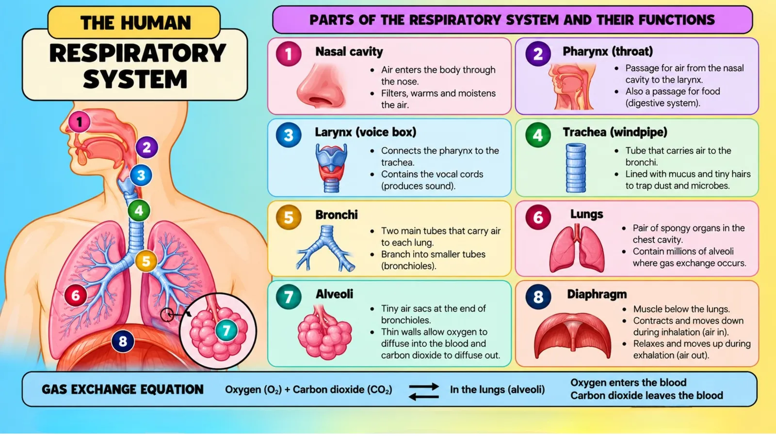
First, the nasal cavity is lined with tiny hairs and sticky mucus that trap dust, pollen and bacteria before they can reach the lungs. Second, the rich blood supply in the nasal lining warms incoming air to body temperature, so freezing air never reaches the delicate lung tissue. Third, the moist lining adds water vapour to the air, and moist air is far gentler on the alveoli than dry air.
Mouth breathing skips all three of these services, which is why people who habitually breathe through the mouth often have drier throats and catch more throat infections.
The Pharynx and Larynx
Air passes from the nasal cavity into the pharynx, the shared passage at the back of the throat. From here it enters the larynx, commonly called the voice box.
The larynx contains the vocal cords, two folds of tissue that vibrate as air passes over them to produce sound. Tightening the cords raises the pitch of your voice and loosening them lowers it. During puberty the larynx grows larger in males, which lengthens the vocal cords and deepens the voice.
Guarding the entrance to the larynx is the epiglottis, a flap of cartilage that folds down whenever you swallow so that food goes into the food pipe rather than the airway. When this flap is even slightly late, food enters the wrong tube and you cough hard until it is cleared.
The Trachea
The trachea, or windpipe, is a tube roughly twelve centimetres long that carries air down towards the lungs. Its most noticeable feature is a series of C shaped rings of cartilage along its length.
These rings exist to keep the tube permanently open. Unlike the food pipe, which stays flattened until food passes through, the airway must never collapse, because air must be able to move at all times. The rings are incomplete at the back, forming a C rather than a full circle, which leaves a soft region where the oesophagus can bulge slightly as food passes. It is an elegant piece of design that solves two problems at once.
The trachea is also lined with ciliated cells and mucus producing cells. The mucus traps particles and the cilia, which are microscopic hair like structures, beat steadily upwards to sweep the trapped dirt towards the throat where it is swallowed or coughed out. This cleaning conveyor belt is often called the mucociliary escalator.
The Bronchi and Bronchioles
At its lower end the trachea divides into two tubes called bronchi, one entering each lung. Inside the lung, each bronchus divides again and again into smaller and smaller tubes called bronchioles, much like the branches of a tree becoming twigs.
This branching structure is the reason the lungs are sometimes described as a respiratory tree. As the tubes get smaller, cartilage disappears and smooth muscle takes over. That smooth muscle can tighten or relax, changing the width of the airway. In asthma, this muscle tightens too much and the airways narrow, which makes breathing difficult.
The Alveoli: Where the Real Work Happens
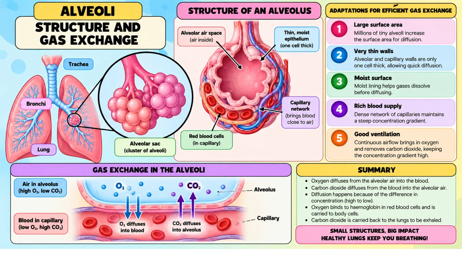
At the end of the smallest bronchioles sit clusters of tiny air sacs called alveoli. This is where gas exchange actually takes place, and everything before this point is simply plumbing to deliver air here.
There are roughly three hundred million alveoli in a pair of human lungs. Individually they are microscopic, but together their surface area is around seventy square metres, which is close to the floor area of a small apartment folded into your chest.
Alveoli are superbly adapted for their job.
The wall of an alveolus is only one cell thick, so gases have a very short distance to travel.
Each alveolus is wrapped in a dense network of capillaries, whose walls are also one cell thick.
Their huge combined surface area allows enormous amounts of gas to be exchanged at once.
Their inner surface is moist, and gases must dissolve in liquid before they can diffuse across a membrane.
A constant blood flow carries oxygen away as fast as it arrives, keeping the concentration gradient steep.
The Lungs, Diaphragm and Ribs
The lungs are two spongy, elastic organs in the chest cavity. The right lung has three lobes and the left has two, because the heart occupies some space on the left side. Each lung is covered by two thin membranes called pleura, with a slippery fluid between them that lets the lungs slide smoothly during breathing.
Below the lungs lies the diaphragm, a large dome shaped sheet of muscle separating the chest from the abdomen. Between the ribs lie the intercostal muscles. These two muscle groups do the physical work of breathing, because the lungs themselves contain no muscle and cannot inflate by their own effort.
🧩
Slide: Parts and their functions
Nose to diaphragm, one job each
1/1
The Mechanism of Breathing
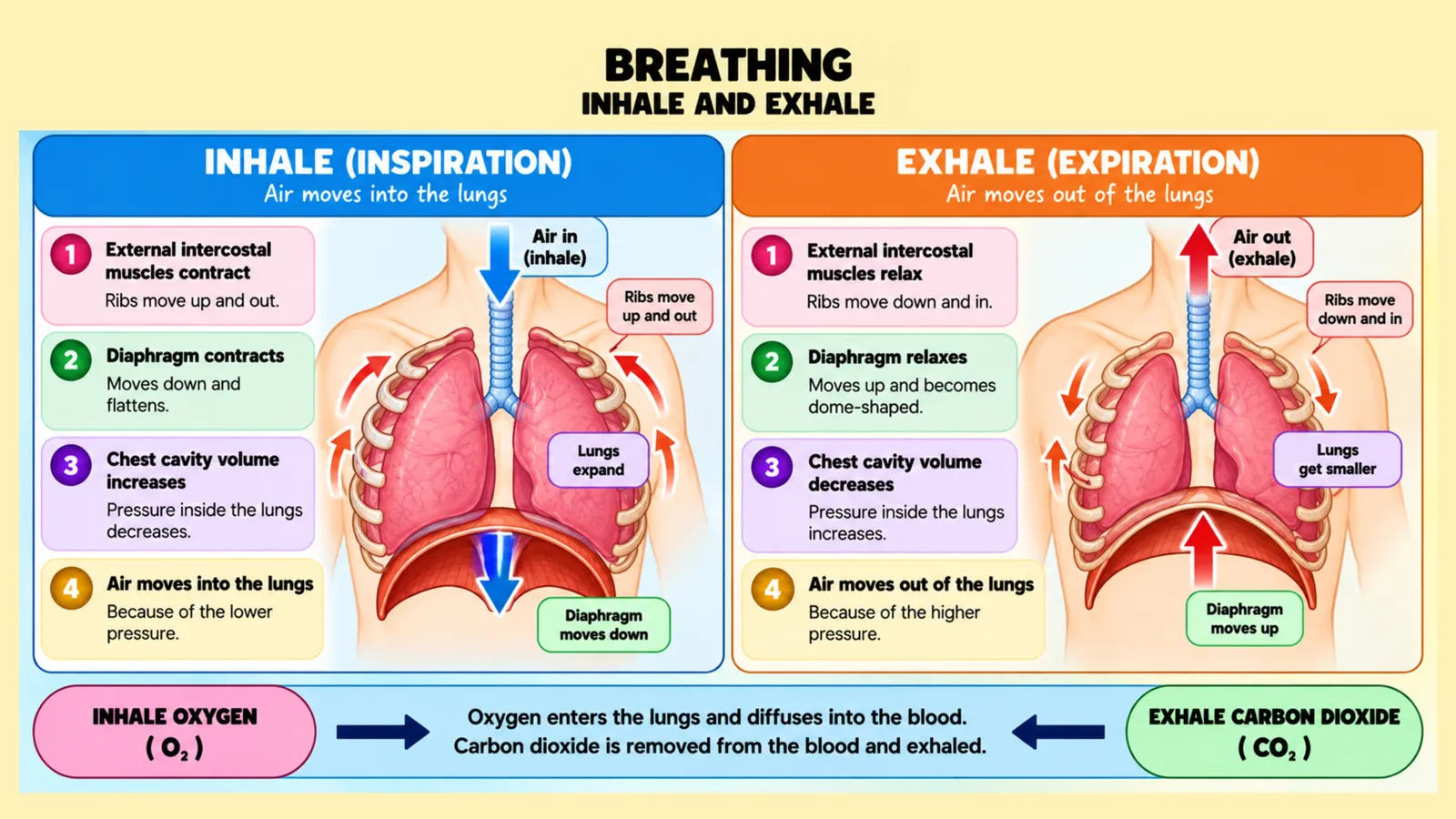
Breathing works on a simple physical principle. Air always moves from a region of higher pressure to a region of lower pressure. Your body does not suck air in. It changes the volume of the chest, which changes the pressure, and air moves accordingly.
Inhalation, or Breathing In
Inhalation is an active process, meaning it requires muscular effort.
The diaphragm contracts and flattens downwards. The external intercostal muscles contract and pull the ribs upwards and outwards. Both movements increase the volume of the chest cavity. Because the same amount of air now occupies a larger space, the pressure inside the lungs falls below the pressure of the outside air. Air therefore rushes in through the nose and down the airways until the pressures are equal.
Exhalation, or Breathing Out
Exhalation during quiet breathing is largely passive, meaning it happens mostly through elastic recoil rather than effort.
The diaphragm relaxes and returns to its dome shape. The intercostal muscles relax and the ribs move down and inwards. The chest volume decreases, the pressure inside the lungs rises above the outside air pressure, and air flows out.
During heavy exercise or forceful blowing, exhalation becomes active as abdominal muscles and internal intercostal muscles push air out more powerfully.
🌬️
Slide: Breathing in and breathing out
Muscles, volume and pressure
1/1
Gas Exchange: How Oxygen Enters the Blood
Gas exchange happens by diffusion, which is the movement of particles from where they are more concentrated to where they are less concentrated. No energy is required, and no pumping is involved. The body simply arranges the concentrations so that diffusion does the work.
Air arriving in an alveolus is rich in oxygen. The blood arriving in the surrounding capillary has come from the body tissues and is low in oxygen and high in carbon dioxide. Oxygen therefore diffuses from the alveolus into the blood, and carbon dioxide diffuses from the blood into the alveolus to be breathed out.
Inside the blood, oxygen is carried by haemoglobin, the red pigment inside red blood cells. Haemoglobin binds oxygen where oxygen is plentiful, forming oxyhaemoglobin, and releases it where oxygen is scarce, which is exactly what happens in working tissues. This is why blood leaving the lungs is bright red and blood returning from the body is a darker shade.
Carbon dioxide is carried differently. Most of it travels dissolved in the plasma as bicarbonate ions, some binds to haemoglobin, and a small amount dissolves directly in the plasma.
🫧
Slide: Alveoli and gas exchange
Why the air sacs are so good at their job
1/1
Composition of Inhaled and Exhaled Air
Inhaled air contains about twenty one percent oxygen, while exhaled air contains about sixteen percent. Your body does not use all the oxygen it takes in.
Inhaled air contains about zero point zero four percent carbon dioxide, while exhaled air contains about four percent, roughly one hundred times more.
Nitrogen makes up about seventy eight percent of both, because the body does not use it.
Exhaled air is warmer and much more saturated with water vapour, which is why your breath is visible on a cold morning.
The fact that exhaled air still contains sixteen percent oxygen is the scientific reason rescue breathing during first aid actually works.
Lung Volumes You Should Know
Tidal volume is the amount of air moved in one normal quiet breath, roughly five hundred millilitres.
Inspiratory reserve volume is the extra air you can force in after a normal breath in.
Expiratory reserve volume is the extra air you can force out after a normal breath out.
Residual volume is the air that always stays in the lungs, which prevents the alveoli from collapsing completely.
Vital capacity is the largest volume you can breathe out after the deepest possible breath in, and it is often used to assess lung health.
Common Respiratory Diseases
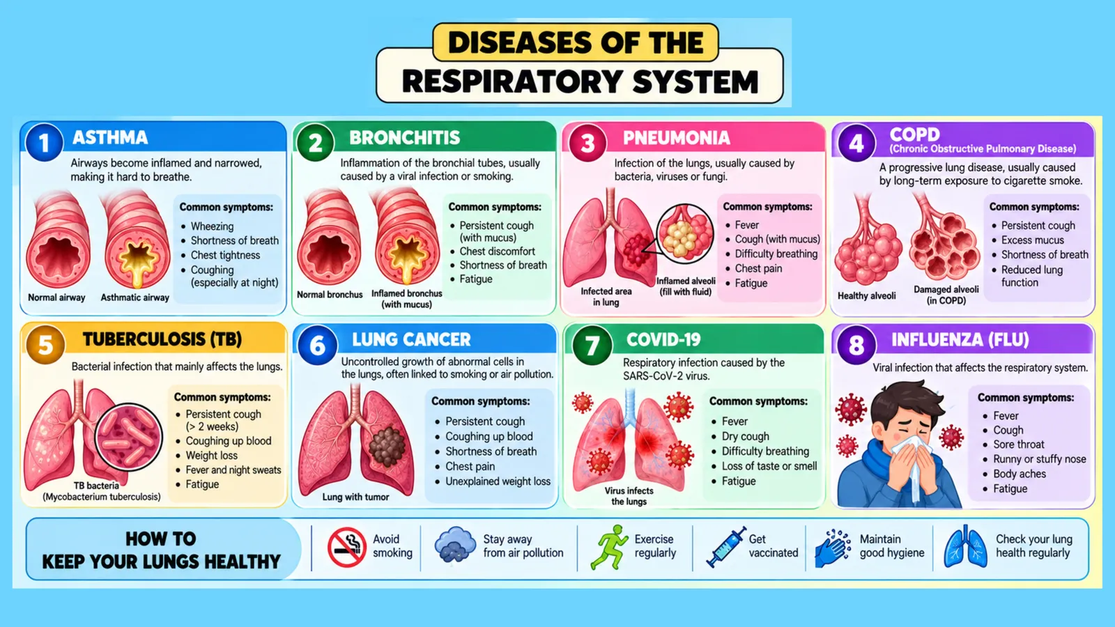
Asthma involves narrowed, inflamed airways that cause wheezing and difficulty breathing, often triggered by dust, cold air, pollen or exercise.
Bronchitis is inflammation of the bronchi with excess mucus, producing a persistent cough.
Pneumonia is an infection in which alveoli fill with fluid, reducing the surface available for gas exchange.
Emphysema involves destruction of alveolar walls so that many small sacs merge into fewer large ones, drastically reducing surface area. Smoking is the leading cause.
Tuberculosis is a bacterial infection that damages lung tissue and remains a serious public health problem in many regions.
🩺
Slide: Diseases of the respiratory system
Causes, symptoms and keeping your lungs healthy
1/1
How Smoking Damages the Respiratory System
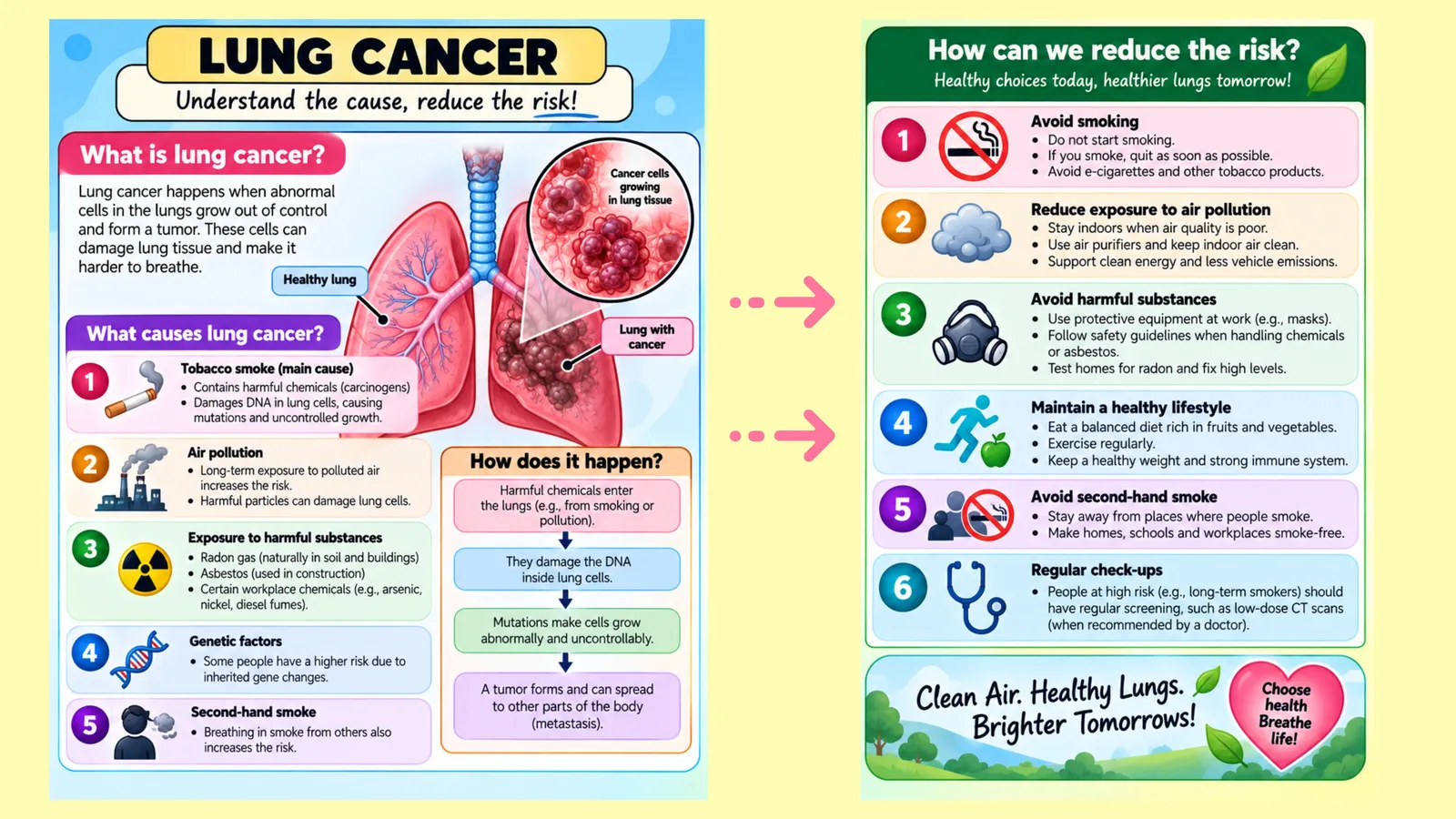
Smoking harms the system at almost every level. Tar coats the airways and paralyses the cilia, so trapped dirt can no longer be swept out and the smoker develops a persistent cough. Chemicals in smoke inflame the bronchi and increase mucus production. Over years, the delicate alveolar walls break down, permanently reducing surface area. Carbon monoxide in the smoke binds to haemoglobin far more strongly than oxygen does, so less oxygen can be carried in the blood. Many of the compounds in tobacco smoke are also carcinogens that directly damage the genetic material of lung cells.
🚭
Slide: Lung cancer
Understand the causes, reduce the risk
1/1
Frequently Asked Questions About the Respiratory System
What is the difference between breathing and respiration?
Breathing is the movement of air in and out of the lungs. Respiration is the chemical release of energy from glucose inside cells. Breathing supplies the oxygen that respiration requires.
Why are the tracheal cartilage rings shaped like a C instead of a full circle?
The cartilage keeps the airway permanently open, and the gap at the back allows the food pipe behind it to expand when food passes down.
Why do we breathe faster during exercise?
Working muscles use more oxygen and release more carbon dioxide. Sensors detect the rising carbon dioxide level in the blood and increase both the rate and depth of breathing to correct it.
Which gas actually controls the urge to breathe?
Carbon dioxide. Rising carbon dioxide levels in the blood are the main trigger for increased breathing, not falling oxygen levels.
Why does the left lung have only two lobes?
The heart is positioned slightly to the left, so the left lung has a notch that gives the heart room, leaving space for only two lobes instead of three.
Final Thoughts
The respiratory system is a beautiful example of biology solving a physics problem. The body needs to move an enormous quantity of gas across a membrane, so it builds three hundred million tiny sacs to create a surface area larger than a room, keeps their walls a single cell thick, wraps them in blood vessels, and then uses simple pressure changes to pump air in and out.
When revising, focus on two things above all. Be able to describe the exact muscle movements of inhalation and exhalation in order, and be able to list the adaptations of the alveoli and explain why each one helps. Those two answers appear in examinations year after year, and both are far easier to remember once you understand the reasoning behind them rather than the wording alone.
🗂️ Revision Flashcards
Tap a card to reveal the answer.
🎯 Quick Quiz
8 questions. Pick an answer to check it straight away.
1Where does respiration take place?
Respiration is the chemical process in the mitochondria of every living cell; breathing happens in the chest.
2What happens to the diaphragm during inhalation?
The diaphragm contracts and flattens, increasing chest volume so pressure falls and air flows in.
3By what process does oxygen move from the alveoli into the blood?
Gas exchange happens by diffusion from high to low concentration, needing no energy.
4About how much oxygen does exhaled air still contain?
Exhaled air holds about 16% oxygen, which is why rescue breathing works.
5Which gas is the main trigger for the urge to breathe?
Rising carbon dioxide levels in the blood mainly trigger breathing, not falling oxygen.
6Why does the left lung have only two lobes?
The heart sits slightly to the left, so the left lung has room for only two lobes.
7Which disease destroys alveolar walls so small sacs merge into fewer large ones?
Emphysema drastically reduces surface area; smoking is the leading cause.
8How does carbon monoxide in smoke reduce oxygen transport?
Carbon monoxide binds haemoglobin far more strongly than oxygen, so less oxygen is carried.