Prokaryotes were the first life forms on Earth, appearing around 3.5 billion years ago, and they are still the most numerous. There are more bacterial cells in a single teaspoon of garden soil than there are people on the planet. Prokaryotes live in boiling hot springs, frozen Antarctic ice, the deepest ocean trenches and inside your own gut. Some cause deadly diseases, while others make oxygen, recycle nutrients and help you digest food. In this guide you will learn what prokaryotic cells are, what they look like inside, how they reproduce and how they differ from the eukaryotic cells that make up your body.
📖 Lesson
Structure, size, shapes, Gram staining, binary fission and how prokaryotes differ from eukaryotic cells.
What Is a Prokaryotic Cell?
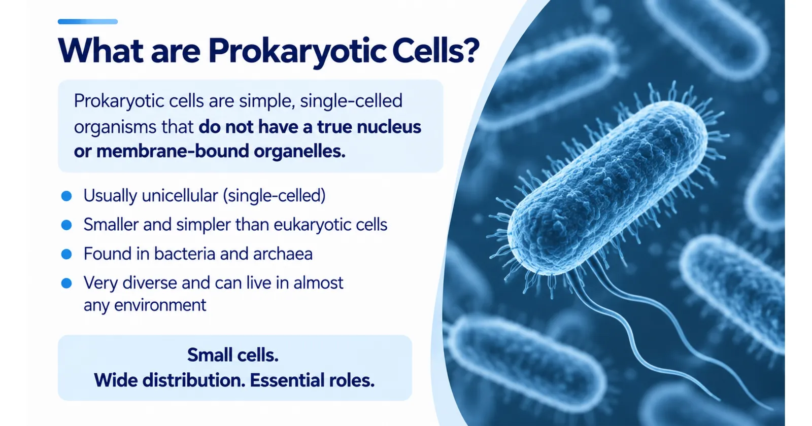
The word prokaryote comes from the Greek words "pro", meaning before, and "karyon", meaning kernel or nucleus. Prokaryotes are cells that existed before the nucleus evolved.
The key feature to remember is: prokaryotic cells do not have a true nucleus. Their genetic material is not enclosed in a nuclear membrane. They also lack other membrane-bound organelles such as mitochondria, chloroplasts, the endoplasmic reticulum and the Golgi apparatus.
All prokaryotes are unicellular, meaning each organism is a single cell, although some form chains, clusters or colonies.
Slides: What are prokaryotic cells?
Simple cells, big impact
1/2
How Big Are Prokaryotic Cells?
Prokaryotic cells are tiny, usually between 0.1 and 5 micrometres (µm) long. A micrometre is a thousandth of a millimetre. Most eukaryotic cells are 10 to 100 µm across, so a typical bacterium is around 10 times smaller than a human cell. You need a light microscope at high magnification to see bacteria, and an electron microscope to see their internal structures.
Their small size gives prokaryotes a large surface area to volume ratio, which allows nutrients and waste to diffuse in and out quickly. This is one reason bacteria can grow and divide so fast.
Structure of a Prokaryotic Cell: Parts and Functions
A typical bacterial cell contains some structures found in all prokaryotes and some that only certain species have.
Structures found in all prokaryotic cells
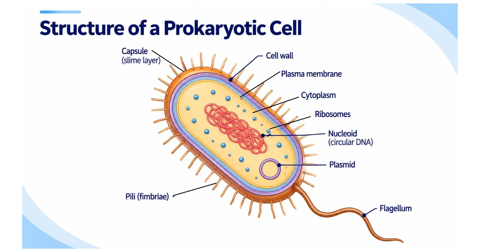
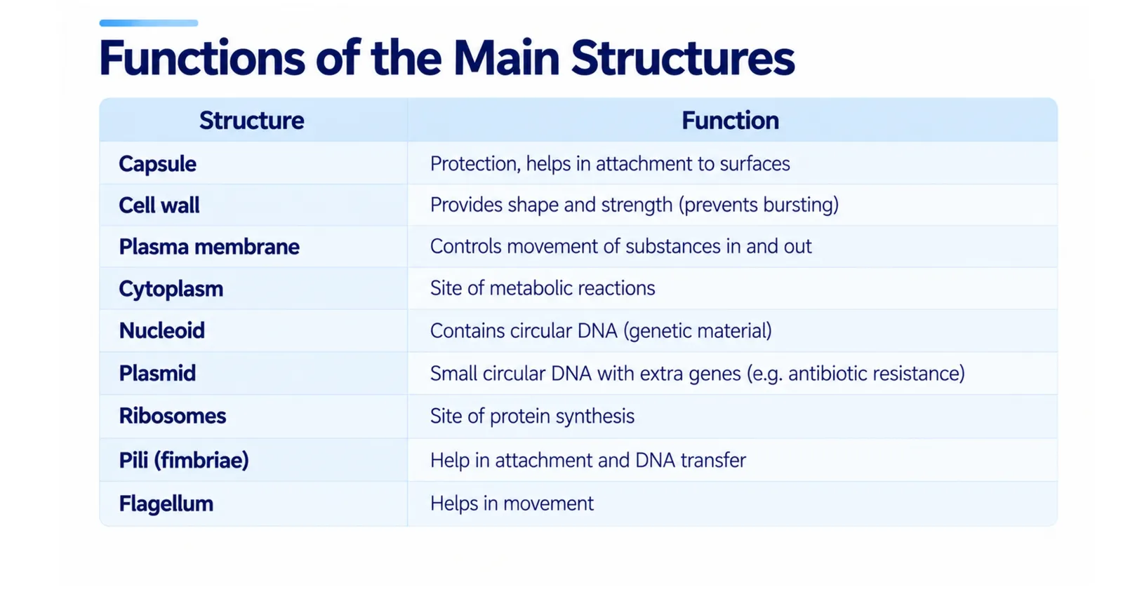
Cell membrane (plasma membrane): a partially permeable membrane that controls which substances enter and leave the cell. In prokaryotes, the membrane is also where respiration takes place, because there are no mitochondria.
Cytoplasm: a jelly-like fluid where most chemical reactions happen. It contains enzymes, nutrients, ribosomes and the DNA.
Nucleoid (circular DNA): the region of cytoplasm containing the main chromosome, a single, large loop of DNA that carries the genes needed for the cell to survive and reproduce. It is not surrounded by a membrane.
Ribosomes (70S): tiny structures that make proteins by protein synthesis. Prokaryotic ribosomes are smaller than eukaryotic ones, which are 80S. The "S" stands for Svedberg units, a measure of size based on how fast particles settle in a centrifuge.
Structures found in most prokaryotic cells
- Cell wall: a rigid layer outside the membrane that gives the cell its shape and stops it bursting when water enters by osmosis. In bacteria it is made of peptidoglycan (also called murein), not cellulose as in plants or chitin as in fungi.
Structures found in some prokaryotic cells
Plasmids: small, separate rings of DNA. They carry extra genes that are useful but not essential, such as genes for antibiotic resistance. Plasmids can copy themselves independently and can be passed between bacteria. Scientists use plasmids as vectors in genetic engineering, for example to insert the human insulin gene into bacteria.
Capsule (slime layer): a sticky outer layer of polysaccharides that protects the cell from drying out and from being engulfed by white blood cells. It also helps bacteria stick to surfaces. Capsules make some bacteria more dangerous.
Flagellum (plural flagella): a long, whip-like tail that rotates like a propeller to move the cell through liquid.
Pili (singular pilus): short, hair-like projections that help bacteria attach to surfaces and to other cells. Special sex pili are used to transfer plasmids between bacteria in a process called conjugation.
Mesosomes: infoldings of the membrane seen in older electron micrographs. Scientists now think they are artefacts created when samples are prepared, so they are no longer considered true structures.
Slides: Structure of a prokaryotic cell
Labelled bacterium and what each part does
1/2
Prokaryotic vs Eukaryotic Cells: Key Differences
This comparison is one of the most common exam questions in cell biology.
| Feature | Prokaryotic cells | Eukaryotic cells |
|---|---|---|
| Examples | Bacteria and archaea | Animals, plants, fungi, protists |
| Nucleus | No true nucleus; DNA in nucleoid | True nucleus with a nuclear envelope |
| DNA form | Circular, not associated with histone proteins (in bacteria) | Linear chromosomes wrapped around histones |
| Plasmids | Often present | Rare (found in some yeasts) |
| Membrane-bound organelles | Absent | Present (mitochondria, ER, Golgi, chloroplasts in plants) |
| Ribosomes | Smaller, 70S | Larger, 80S in the cytoplasm |
| Cell wall | Usually present, made of peptidoglycan in bacteria | Cellulose in plants, chitin in fungi, absent in animals |
| Size | Usually 0.1 to 5 µm | Usually 10 to 100 µm |
| Cell division | Binary fission | Mitosis and meiosis |
| Organisation | Always unicellular | Unicellular or multicellular |
What prokaryotic and eukaryotic cells have in common
Despite their differences, all cells share certain features: a cell membrane, cytoplasm, ribosomes and DNA as their genetic material. They all use the same genetic code and carry out protein synthesis in a similar way.
Examples of Prokaryotes
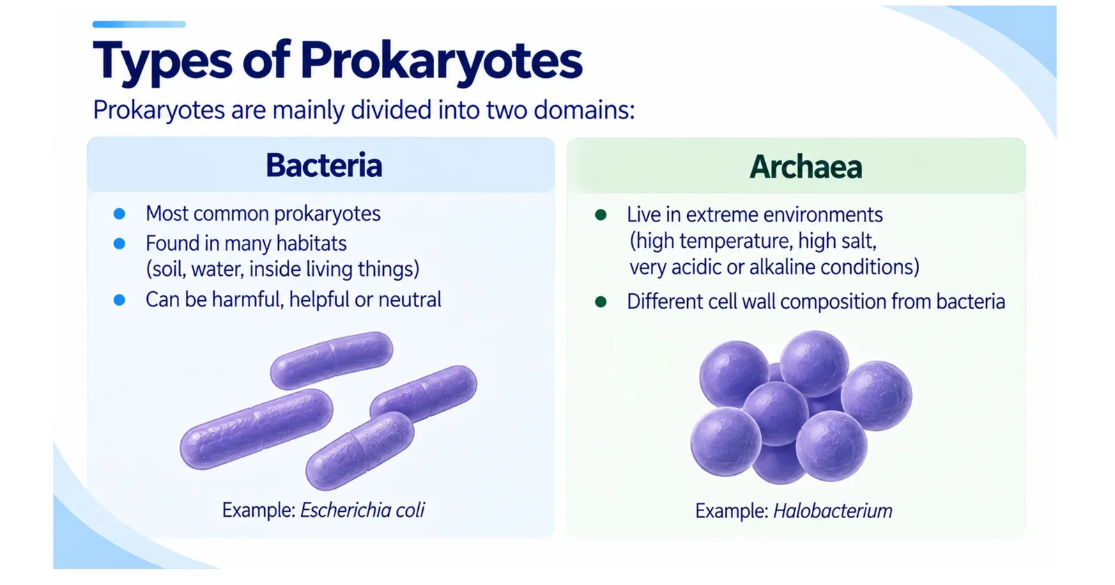
Bacteria
Escherichia coli (E. coli): lives harmlessly in the human gut, although some strains cause food poisoning. It is widely used in laboratories.
Streptococcus: causes strep throat and some forms of pneumonia.
Staphylococcus aureus: found on the skin; can cause infections, including the antibiotic-resistant form MRSA.
Lactobacillus: used to make yoghurt and cheese, and found in probiotic foods.
Mycobacterium tuberculosis: causes tuberculosis (TB).
Cyanobacteria: photosynthetic bacteria that produce oxygen. Ancient cyanobacteria are responsible for filling Earth's atmosphere with oxygen billions of years ago.
Rhizobium: lives in the root nodules of legumes such as peas and beans and fixes nitrogen from the air.
Archaea
Archaea look like bacteria under the microscope, but they are chemically very different. Their cell walls do not contain peptidoglycan, and their membranes are made of unusual lipids. Many are extremophiles that live in harsh environments, such as salty lakes, acidic hot springs and deep-sea hydrothermal vents. Methanogens, archaea that produce methane gas, live in swamps and in the guts of cows. Scientists now think archaea are more closely related to eukaryotes than bacteria are.
Slide: Types of prokaryotes
Bacteria and archaea
1/1
Shapes of Bacteria
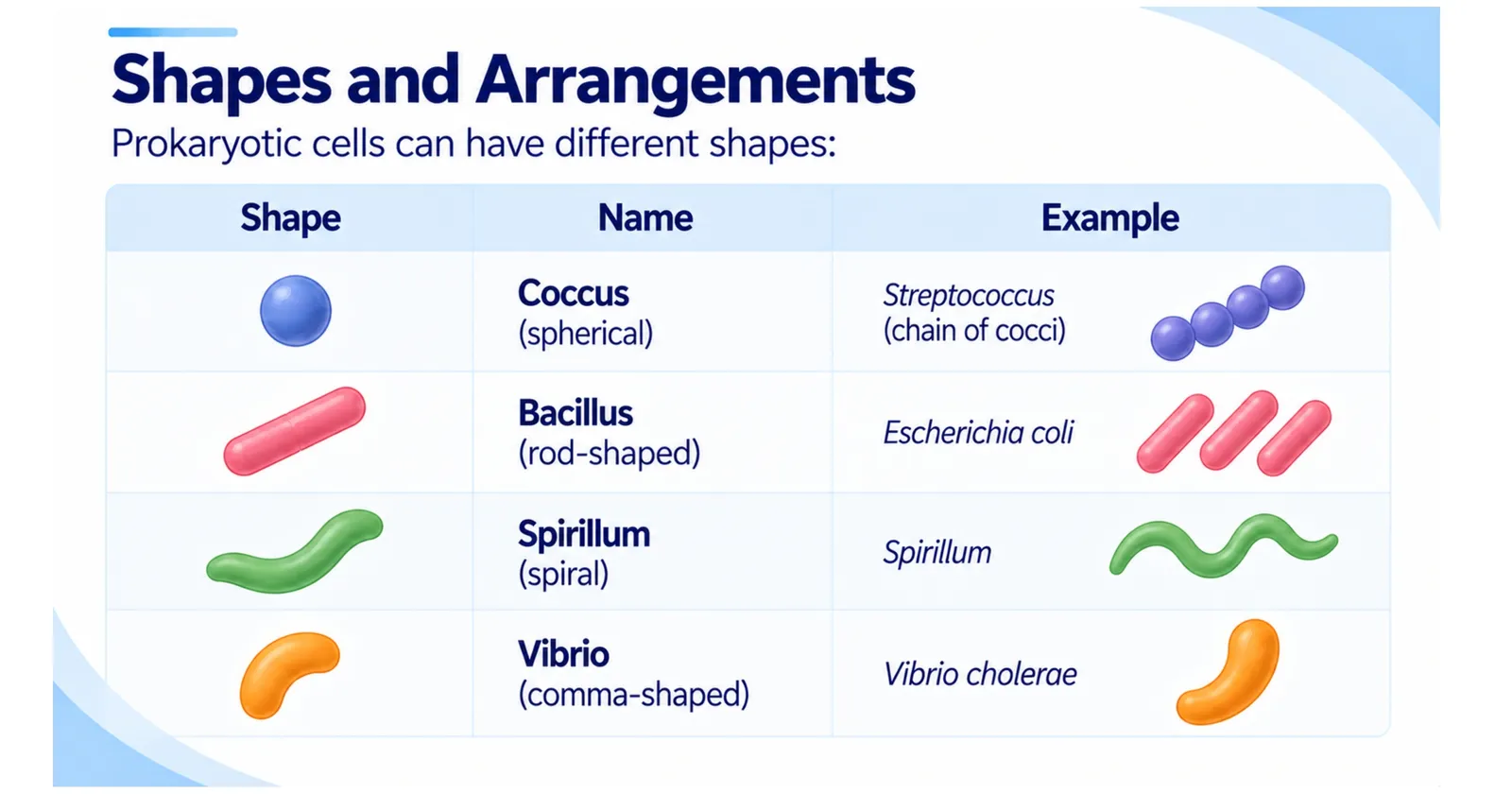
Bacteria are often identified by their shape:
Cocci: spherical, such as Streptococcus (in chains) and Staphylococcus (in clusters).
Bacilli: rod-shaped, such as E. coli.
Spirilla: spiral-shaped, such as the bacterium that causes stomach ulcers.
Vibrios: comma-shaped, such as the bacterium that causes cholera.
Slide: Shapes of bacteria
Coccus, bacillus, spirillum and vibrio
1/1
Gram-Positive and Gram-Negative Bacteria
The Gram stain, developed by Hans Christian Gram in 1884, divides bacteria into two groups based on their cell walls.
Gram-positive bacteria have a thick peptidoglycan wall that holds the purple stain, so they appear purple under the microscope.
Gram-negative bacteria have a thin peptidoglycan layer surrounded by an outer membrane. They lose the purple stain and appear pink or red.
This matters in medicine because the two groups respond differently to antibiotics. For example, penicillin works best against Gram-positive bacteria, because it attacks peptidoglycan synthesis and the outer membrane of Gram-negative bacteria helps block it.
How Do Prokaryotes Reproduce? Binary Fission
Prokaryotes reproduce asexually by binary fission, which means "splitting in two".
The circular DNA is copied.
Plasmids are also copied, sometimes several times.
The cell grows larger, and the two DNA copies move to opposite ends of the cell.
The cell membrane pinches inwards and a new cell wall forms across the middle.
The cell divides into two genetically identical daughter cells.
In ideal conditions, with plenty of nutrients, warmth and moisture, some bacteria can divide every 20 minutes. Starting with one cell, that would produce over a million cells in about seven hours. This explains why food poisoning bacteria multiply so quickly in food left at room temperature.
How bacteria gain new genes
Although binary fission produces clones, bacteria can still share genes through conjugation (passing plasmids through a pilus), transformation (taking up DNA from their surroundings) and transduction (having DNA carried in by viruses). This horizontal gene transfer is one reason antibiotic resistance spreads so quickly.
How Prokaryotes Get Their Food and Energy
Prokaryotes have a greater variety of ways to obtain energy than any other group of living things.
Photoautotrophs, such as cyanobacteria, use light energy to make their own food by photosynthesis. They do not have chloroplasts; instead, their chlorophyll is found on folded internal membranes called thylakoids.
Chemoautotrophs make food using energy released from chemical reactions, for example by oxidising ammonia, iron or sulfur compounds. Some live around deep-sea vents where no sunlight reaches, forming the base of entire food chains.
Heterotrophs, which include most bacteria, feed on organic matter. Saprotrophic bacteria digest dead material, while parasitic bacteria feed on living hosts.
Prokaryotes also vary in how they use oxygen. Obligate aerobes need oxygen for respiration, obligate anaerobes are poisoned by it and facultative anaerobes such as E. coli can live with or without oxygen.
Surviving tough times: endospores
Some bacteria, such as those that cause tetanus and anthrax, can form endospores when conditions become harsh. An endospore is a tough, dormant structure with a thick protective coat that contains a copy of the DNA. Endospores can survive boiling, freezing, drying and many disinfectants for years, and even centuries, and then germinate into active bacteria when conditions improve. This is why surgical instruments must be sterilised at high temperature and pressure in an autoclave, and why canned food must be heated thoroughly during processing to destroy the spores of dangerous food-poisoning bacteria.
Why Prokaryotes Matter: Good and Bad
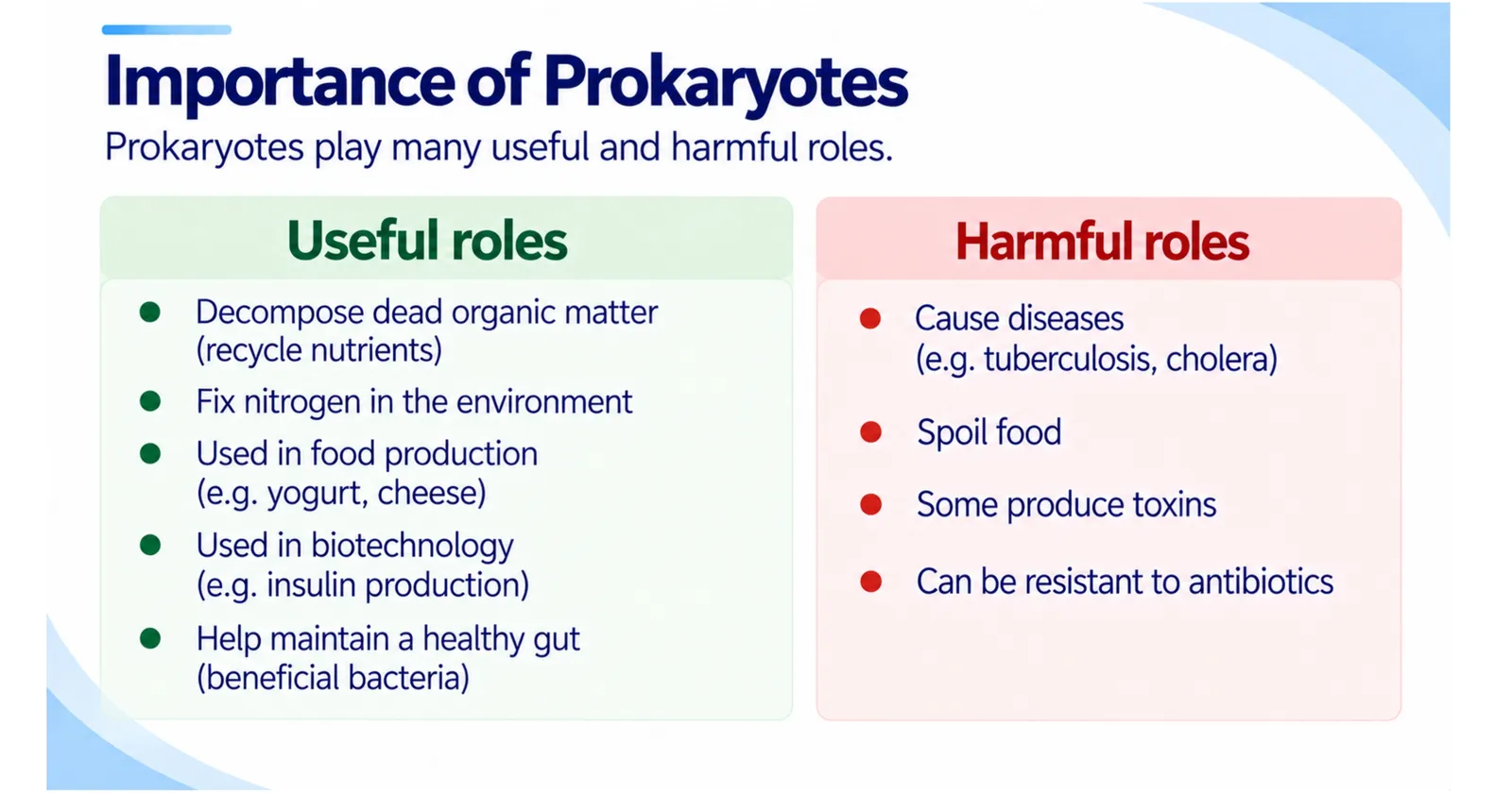
Helpful prokaryotes
Gut bacteria help digest food and make vitamin K.
Decomposer bacteria recycle nutrients by breaking down dead organisms.
Nitrogen-fixing bacteria make nitrogen available to plants.
Bacteria are used to make yoghurt, cheese, vinegar and many medicines.
Genetically modified bacteria produce insulin and other life-saving drugs.
Harmful prokaryotes
Pathogenic bacteria cause diseases such as tuberculosis, cholera, typhoid, tetanus and some types of pneumonia. Many produce toxins that damage cells. Antibiotics can kill bacteria or stop them growing, often by targeting prokaryote-specific structures such as peptidoglycan cell walls and 70S ribosomes. Overusing antibiotics has led to resistant strains, which is a major global health threat.
Slide: Importance of prokaryotes
Useful and harmful roles
1/1
Is a Virus a Prokaryote?
No. Viruses are not cells at all. They have no cytoplasm, no ribosomes and no cell membrane, and they can reproduce only inside a host cell. This is why antibiotics do not work against viral infections such as colds and flu.
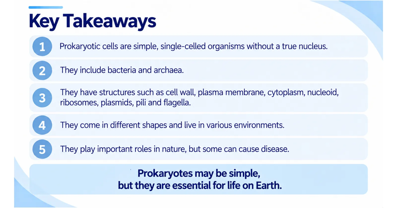
Slide: Key takeaways
1/1
Frequently Asked Questions About Prokaryotic Cells
Do prokaryotes have mitochondria?
No. Prokaryotes carry out respiration on their cell membrane. Interestingly, mitochondria and chloroplasts in eukaryotic cells are thought to have evolved from free-living bacteria that were engulfed by larger cells, an idea called the endosymbiotic theory.
Do prokaryotic cells have DNA?
Yes. They have a single circular chromosome in the nucleoid, and often extra DNA in plasmids.
Are prokaryotes older than eukaryotes?
Yes. Prokaryotes appeared about 3.5 billion years ago, while eukaryotes evolved around 2 billion years ago.
🗂️ Revision Flashcards
Tap a card to reveal the answer.
🎯 Quick Quiz
8 questions. Pick an answer to check it straight away.
1Which feature do prokaryotic cells lack?
Prokaryotic DNA is not enclosed in a nuclear membrane; it sits in the nucleoid.
2Where does respiration take place in a prokaryotic cell?
Prokaryotes have no mitochondria, so respiration happens on the cell membrane.
3What is the bacterial cell wall made of?
Bacterial walls are made of peptidoglycan (murein); cellulose is in plants and chitin in fungi.
4What size are prokaryotic ribosomes?
Prokaryotic ribosomes are 70S, smaller than the 80S ribosomes of eukaryotes.
5A rod-shaped bacterium such as E. coli is called a…
Bacilli are rod-shaped; cocci are spherical, spirilla spiral and vibrios comma-shaped.
6Why do Gram-negative bacteria appear pink or red after a Gram stain?
Their thin peptidoglycan layer is surrounded by an outer membrane, so they lose the purple stain.
7In ideal conditions some bacteria divide every 20 minutes. Roughly how long would one cell take to produce over a million cells?
Dividing every 20 minutes, one cell produces over a million cells in about seven hours.
8Why do antibiotics not work against colds and flu?
Viruses have no cytoplasm, ribosomes or cell membrane, so antibiotics do not work against them.