Your eyes let you read these words, recognise the faces of people you love, judge the speed of a passing car and enjoy millions of shades of colour. Scientists estimate that around 80 percent of the information we receive about the world comes through our eyes. Yet each eyeball is only about 2.4 centimetres across, roughly the size of a large grape. In this guide we explore the structure of the human eye, explain step by step how we see, uncover how the eye focuses and adjusts to light, and look at the most common eye problems and how they are corrected.
📖 Lesson
The structure of the eye, how we see, rods and cones, the pupil reflex, accommodation and how eye defects are corrected.
The Structure of the Human Eye
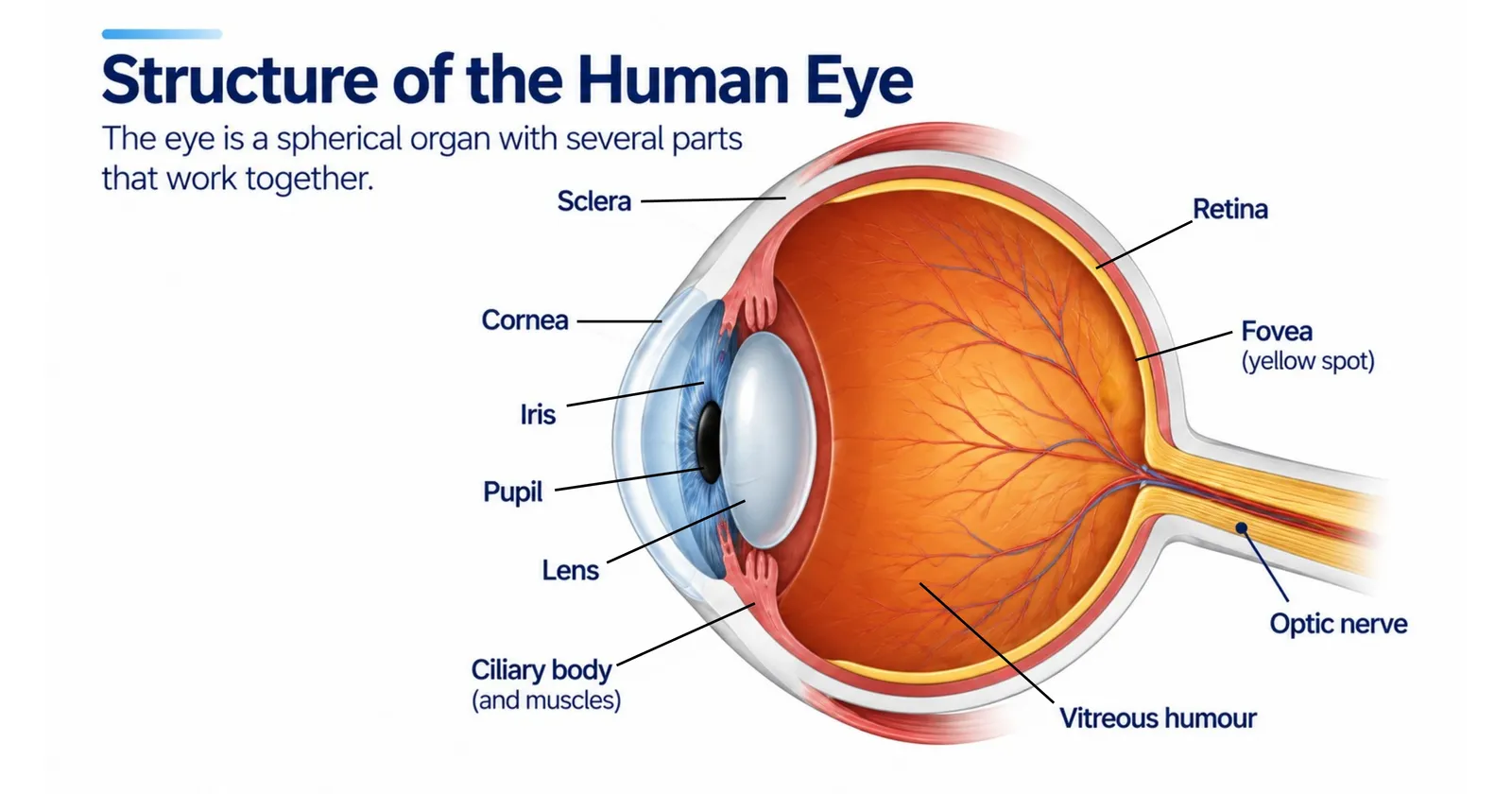
The eyeball sits in a bony socket called the orbit, which protects it. Six muscles attached to the outside of each eyeball move it in every direction. The eyelids, eyelashes and tears protect the front surface.
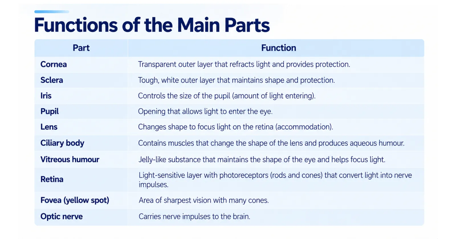
Parts of the eye and their functions
| Part | Description | Function |
|---|---|---|
| Sclera | Tough white outer layer | Protects the eye and gives it shape; muscles attach to it |
| Cornea | Transparent, curved front part of the sclera | Lets light in and does most of the refraction (bending) of light |
| Conjunctiva | Thin transparent membrane over the front of the eye | Protects the cornea and keeps the eye moist |
| Iris | Coloured ring of muscle | Controls the size of the pupil and therefore how much light enters |
| Pupil | Hole in the centre of the iris | Lets light into the eye |
| Lens | Transparent, flexible, biconvex structure | Fine-tunes focusing of light onto the retina |
| Ciliary muscles | Ring of muscle around the lens | Change the shape of the lens for focusing |
| Suspensory ligaments | Fibres connecting the lens to the ciliary muscles | Hold the lens in place |
| Aqueous humour | Watery fluid in front of the lens | Nourishes the cornea and lens and maintains pressure |
| Vitreous humour | Clear jelly filling most of the eyeball | Keeps the eyeball's shape |
| Choroid | Dark layer rich in blood vessels | Supplies nutrients and absorbs stray light to prevent reflections |
| Retina | Light-sensitive layer at the back | Contains rods and cones that detect light |
| Fovea (yellow spot) | Small central pit in the retina | Area of sharpest, most detailed vision; packed with cones |
| Optic nerve | Bundle of nerve fibres | Carries nerve impulses from the retina to the brain |
| Blind spot | Point where the optic nerve leaves the eye | Has no rods or cones, so it cannot detect light |
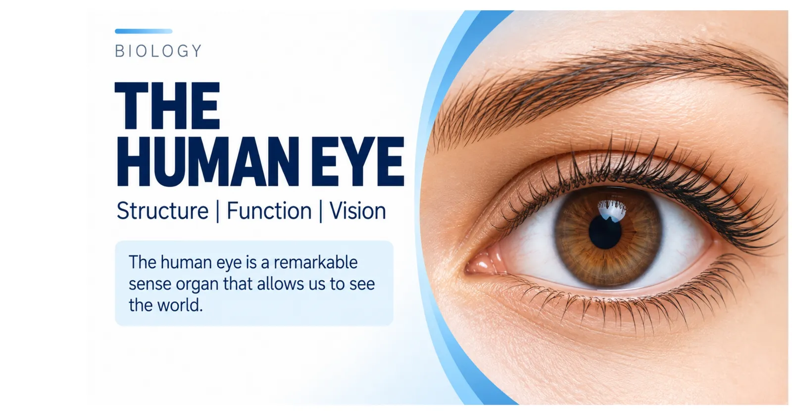
Slides: Structure of the human eye
The parts of the eye and what each one does
1/3
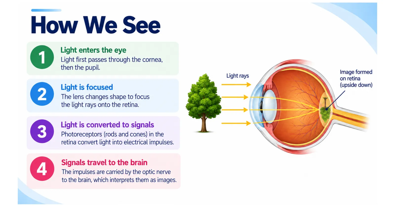
How Do We See? The Process Step by Step
Light reflected from an object enters the eye through the cornea.
The cornea refracts (bends) the light towards the pupil.
Light passes through the aqueous humour and the pupil.
The lens refracts the light further, focusing it through the vitreous humour onto the retina.
An image forms on the retina. It is upside down (inverted), reversed and smaller than the object.
Rods and cones absorb the light and generate electrical nerve impulses.
The optic nerve carries the impulses to the visual cortex at the back of the brain.
The brain interprets the signals, turning the image the right way up and combining the images from both eyes.
Many people are surprised to learn that the cornea, not the lens, does about two-thirds of the focusing. The lens provides the adjustable fine focus.
Slide: How we see
Light enters, is focused, converted to signals and sent to the brain
1/1
Rods and Cones: The Eye's Light Detectors
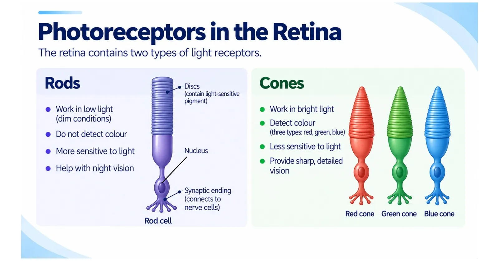
The retina contains two types of photoreceptor cells.
| Feature | Rods | Cones |
|---|---|---|
| Number per eye | About 120 million | About 6 million |
| Sensitivity | Very sensitive to dim light | Need bright light |
| Colour vision | No, only shades of grey | Yes |
| Detail | Low detail | High detail and sharpness |
| Location | Mostly around the edges of the retina | Concentrated in the fovea |
How colour vision works
There are three types of cone, each containing a pigment most sensitive to a different range of wavelengths: red, green or blue light. The brain compares how strongly each type is stimulated to work out colour. Yellow light, for example, stimulates red and green cones. Together, the three cone types allow humans to distinguish up to around a million different colours.
Colour blindness
If one type of cone is missing or does not work properly, a person has colour vision deficiency. The most common form is red-green colour blindness. Because the genes for red and green cone pigments are on the X chromosome, colour blindness is far more common in males, affecting roughly 1 in 12 men but only about 1 in 200 women.
Why we see poorly in the dark
In dim light, cones do not work well, so we rely on rods. That is why it is hard to see colours at night, and why faint stars are easier to see when you look slightly to one side of them, making their light fall on rod-rich areas. When you walk into a dark room, it takes up to 30 minutes for your rods to become fully adapted.
Slide: Rods and cones
The two types of light receptor in the retina
1/1
Controlling Light: The Pupil Reflex
Too much light can damage the retina, while too little makes it hard to see. The iris adjusts the size of the pupil automatically in a reflex action.
The iris contains two sets of muscles: circular muscles arranged in rings and radial muscles arranged like the spokes of a wheel.
In bright light
The circular muscles contract.
The radial muscles relax.
The pupil becomes smaller (constricts), letting less light in and protecting the retina.
In dim light
The circular muscles relax.
The radial muscles contract.
The pupil becomes larger (dilates), letting more light in so that you can see.
This is a reflex, so it is fast and automatic and does not involve conscious thought. Doctors shine a light into the eye to check this reflex after head injuries.
Accommodation: How the Eye Focuses
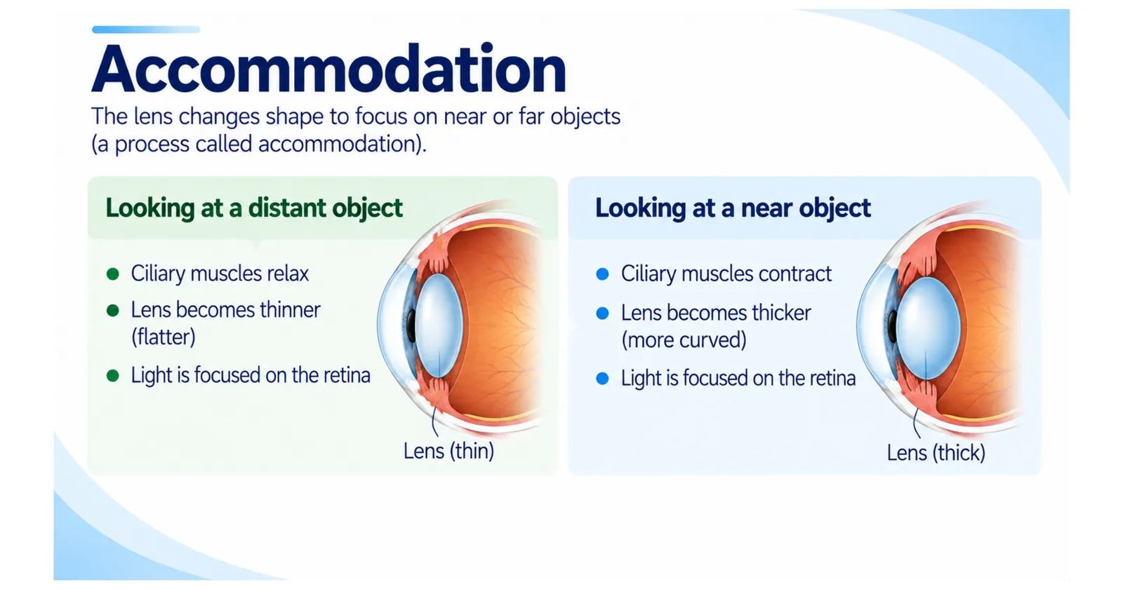
Accommodation is the way the eye changes the shape of the lens to focus on objects at different distances. This is one of the most frequently examined eye topics.
Focusing on a near object
The ciliary muscles contract.
The suspensory ligaments slacken (loosen).
The lens becomes thicker and more rounded (more convex).
Light is refracted more strongly, so it focuses on the retina.
Focusing on a distant object
The ciliary muscles relax.
The suspensory ligaments are pulled tight.
The lens is pulled thinner and flatter.
Light is refracted less, so it focuses on the retina.
A handy memory trick is "near means contract": when you look at something close, your ciliary muscles contract. As we get older, the lens becomes stiffer and cannot become as round, so close reading becomes harder. This is called presbyopia and is why many people need reading glasses after the age of 40 to 45.
Slide: Accommodation
How the lens changes shape for near and distant objects
1/1
The Blind Spot: A Simple Experiment
Where the optic nerve leaves the eye there are no rods or cones, so any light that falls there cannot be detected. You do not usually notice because your brain fills in the gap, and your other eye covers the missing area.
To find your blind spot, draw a cross and a dot about 8 centimetres apart on a card. Close your left eye, stare at the cross with your right eye and slowly move the card towards your face. At a certain distance, the dot disappears. That is the moment its image falls on your blind spot.
Common Eye Defects and How They Are Corrected
Myopia (short-sightedness)
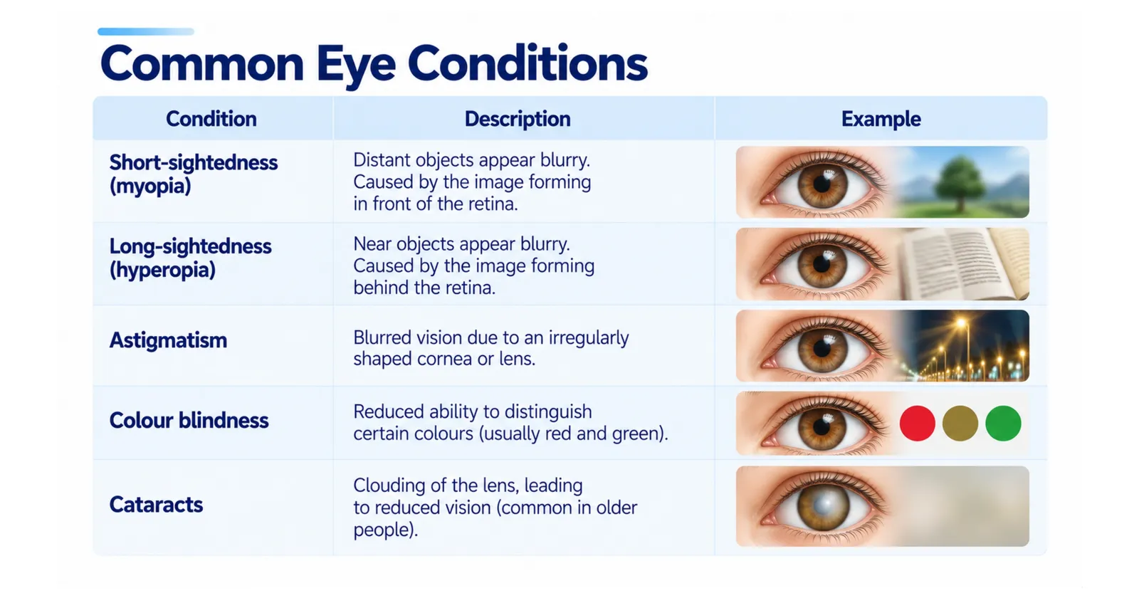
People with myopia can see near objects clearly, but distant objects look blurred. The eyeball is too long or the lens is too curved, so light from distant objects focuses in front of the retina. It is corrected with concave (diverging) lenses, which spread the light out before it enters the eye. Myopia is becoming much more common worldwide, and spending time outdoors in childhood appears to reduce the risk.
Hypermetropia (long-sightedness)
People with hypermetropia can see distant objects clearly, but near objects look blurred. The eyeball is too short or the lens is too weak, so light from near objects would focus behind the retina. It is corrected with convex (converging) lenses.
Astigmatism
An unevenly curved cornea or lens causes blurred or distorted vision at all distances. It is corrected with specially shaped cylindrical lenses.
Cataracts
A cataract is a cloudy area in the lens that makes vision blurry and colours faded. It is common in older people and is treated with a simple operation that replaces the cloudy lens with an artificial one.
Glaucoma
Glaucoma is a build-up of pressure inside the eye that damages the optic nerve. It often has no early symptoms, so regular eye tests are important. It is treated with eye drops, laser treatment or surgery.
Modern treatments
Besides glasses and contact lenses, laser eye surgery can reshape the cornea to correct myopia, hypermetropia and astigmatism. Replacement lenses can also be implanted inside the eye.
Slide: Common eye conditions
Short-sightedness, long-sightedness, astigmatism, colour blindness and cataracts
1/1
Why Two Eyes Are Better Than One
Each eye sees the world from a slightly different angle. The brain combines the two images to judge distance and depth, which is called binocular or stereoscopic vision. This is essential for catching a ball, pouring a drink or crossing a road. Predators such as owls and cats have forward-facing eyes for excellent depth perception, while prey animals such as rabbits have eyes on the sides of their heads, giving them an almost all-round view to spot danger.
Tears and Eye Protection
The eye is protected by several defences:
Eyelids close automatically in the blink reflex to protect against dust and bright light.
Eyelashes trap particles.
Tears, produced by the lacrimal glands, wash the eye, keep the cornea moist and contain lysozyme, an enzyme that kills bacteria.
Eyebrows stop sweat running into the eyes.
We blink about 15 to 20 times every minute, but we blink much less while staring at screens, which can cause dry, tired eyes.
How to Look After Your Eyes
Follow the 20-20-20 rule: every 20 minutes of screen time, look at something 20 feet (about 6 metres) away for 20 seconds.
Wear sunglasses that block UV light.
Eat foods rich in vitamin A, such as carrots, sweet potatoes and leafy greens. Vitamin A is needed to make the pigment in rods, and a deficiency causes night blindness.
Wear safety goggles for sports and practical work.
Have regular eye tests, even if your vision seems fine.
Frequently Asked Questions About the Human Eye
Why is the image on the retina upside down?
The lens is convex, so light rays cross over before reaching the retina. The brain automatically flips the image so we see the world the right way up.
What gives eyes their colour?
Eye colour depends on the amount of melanin in the iris. Brown eyes have lots of melanin; blue eyes have very little and appear blue because of the way light scatters.
Can eyes be transplanted?
A whole eye cannot be transplanted, but the cornea can. Corneal transplants restore sight to thousands of people every year.
Key Takeaways
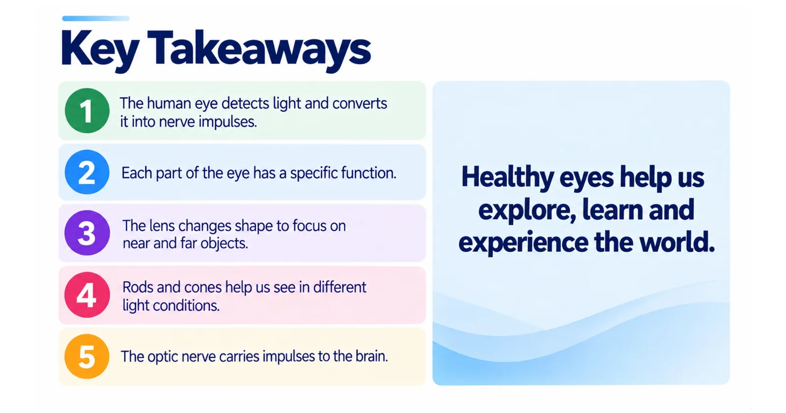
Light is refracted by the cornea and lens and focused onto the retina.
Rods detect dim light; cones detect colour and fine detail.
The iris controls pupil size through a reflex to protect the retina.
Ciliary muscles and suspensory ligaments change the lens shape during accommodation.
Myopia is corrected by concave lenses and hypermetropia by convex lenses.
Slide: Key takeaways
1/1
Your eyes are part of a remarkable network of sense organs. Explore our guides to the human ear and the human nose to complete your understanding of how you sense the world.
🗂️ Revision Flashcards
Tap a card to reveal the answer.
🎯 Quick Quiz
8 questions. Pick an answer to check it straight away.
1Which part of the eye does about two-thirds of the focusing?
The cornea, not the lens, does about two-thirds of the focusing; the lens provides the adjustable fine focus.
2What happens in the iris in bright light?
In bright light the circular muscles contract and the radial muscles relax, so the pupil becomes smaller and protects the retina.
3When the eye focuses on a near object, which statement is correct?
Near means contract: the ciliary muscles contract, the suspensory ligaments slacken and the lens becomes thicker and more convex.
4Myopia (short-sightedness) is corrected with which type of lens?
In myopia light focuses in front of the retina, so concave lenses spread the light out before it enters the eye.
5Which cells in the retina allow us to see colour?
Cones detect colour and fine detail; rods only give shades of grey.
6Why can the blind spot not detect light?
The blind spot is where the optic nerve leaves the eye, and it has no rods or cones.
7What does the lysozyme in tears do?
Tears contain lysozyme, an enzyme that kills bacteria.
8A deficiency of vitamin A causes which condition?
Vitamin A is needed to make the pigment in rods, and a deficiency causes night blindness.