Every organelle's structure and job, how they work together, and the exam phrases to learn.
Introduction: A City Inside Every Cell
Imagine trying to run an entire city inside a space smaller than the full stop at the end of this sentence. The city needs a government, a power station, factories, a road network, a postal service, a recycling department and a boundary wall. Somehow it must fit all of that into a space you cannot see without a microscope.
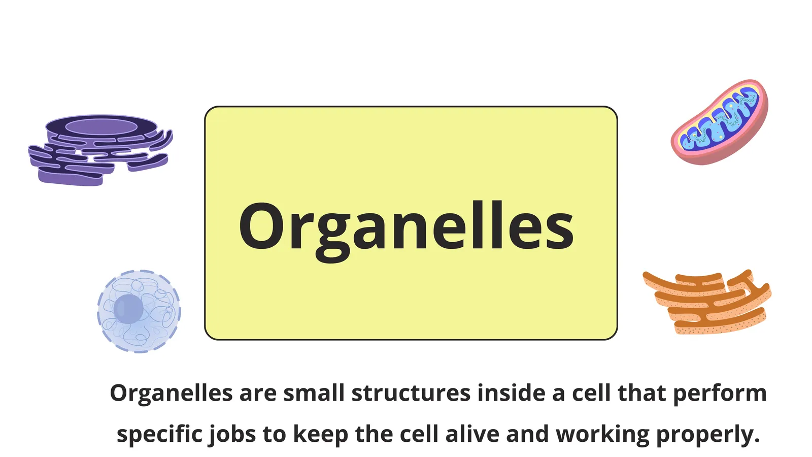
That is exactly what a cell does. The tiny structures inside it are called organelles, and the word itself gives the game away. Organelle means little organ. Just as your body has organs that each do a specific job, a cell has organelles that each do a specific job.
This guide walks through every major organelle, explaining what it looks like, what it does, and why it matters. By the end you should be able to look at a cell diagram and explain not just the names but the reasoning behind the whole arrangement.
🏙️
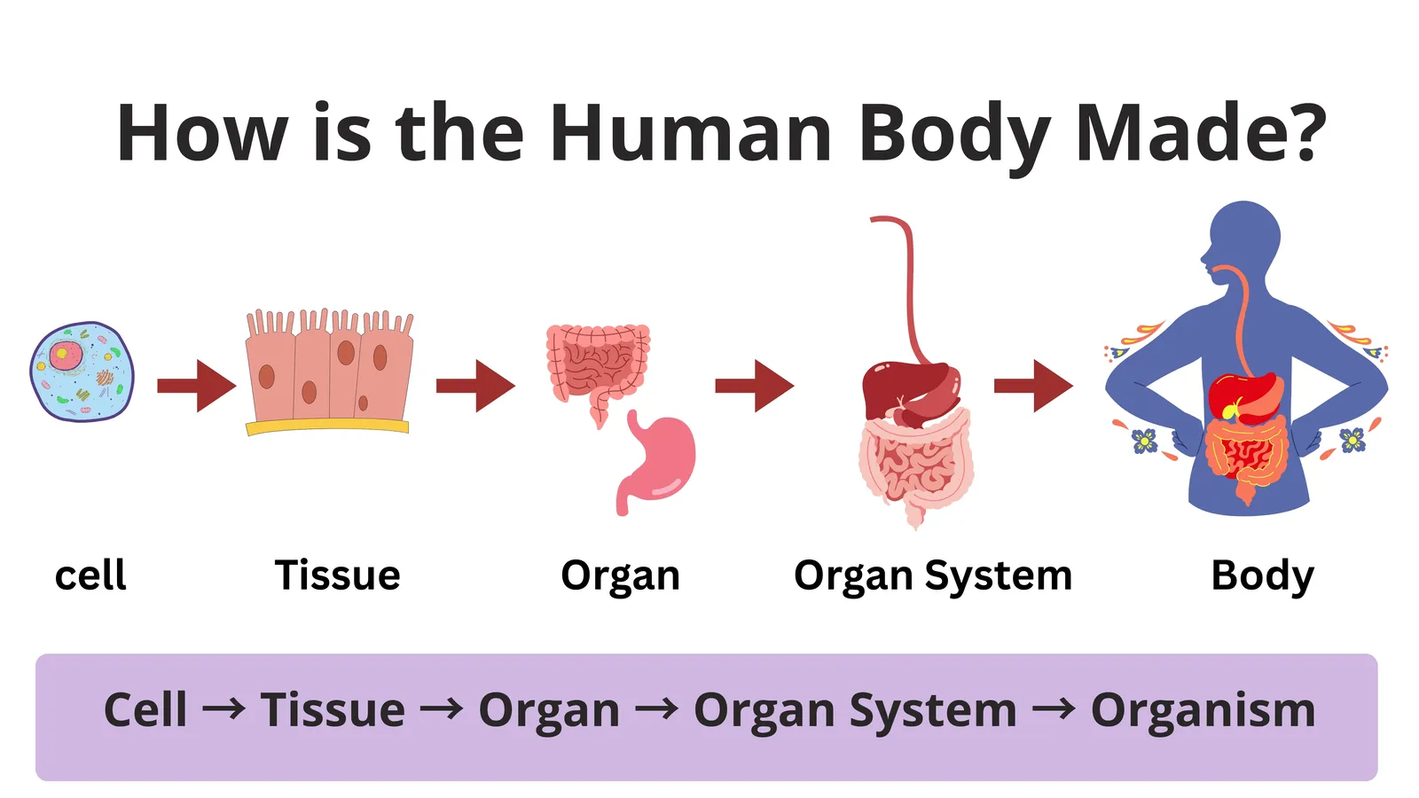
Slide: How is the human body made?
Cells build tissues, organs and organ systems
1/1
What Are Cell Organelles?
An organelle is a specialised structure inside a cell that performs a particular function. Most organelles are surrounded by their own membrane, which keeps their contents separate from the rest of the cytoplasm.
This separation is the whole point. It is called compartmentalisation, and it is one of the most important ideas in cell biology.
Consider why it matters. Lysosomes contain powerful digestive enzymes capable of destroying proteins. If those enzymes floated freely in the cytoplasm they would attack the cell itself. By keeping them sealed inside a membrane bag, the cell can own dangerous tools safely. In the same way, the reactions of respiration need particular conditions that would interfere with other reactions if they happened everywhere at once.
A useful comparison is a school. Chemistry experiments happen in the laboratory, sport happens on the field, and books are kept in the library. Nobody performs a chemistry experiment in the library. Separating activities into rooms makes complex work possible, and organelles are the rooms of the cell.
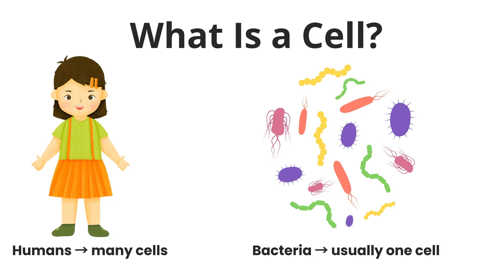
Because prokaryotic cells such as bacteria lack membrane bound organelles, they carry out all their processes in one shared space, which limits how complex they can become.
🔬
Slides: What are organelles?
Little organs with specific jobs
1/2
The Nucleus: The Control Centre
The nucleus is usually the largest and most obvious organelle in a eukaryotic cell. It is typically spherical and is found near the centre of animal cells, while in a mature plant cell the huge vacuole often pushes it to one side.
Structure of the Nucleus
The nucleus is enclosed by a nuclear envelope, which is a double membrane rather than a single one. This envelope is dotted with thousands of nuclear pores, small openings that allow selected molecules to pass in and out. Large DNA molecules stay inside, while messenger molecules and newly built ribosomes leave through the pores.
Inside is a fluid called nucleoplasm containing chromatin, which is DNA wound around proteins. Chromatin looks like a tangled network of threads under a microscope. When the cell prepares to divide, chromatin condenses into distinct rod shaped chromosomes. Human body cells contain forty six of them.
The nucleus also contains at least one dense round region called the nucleolus, which has no membrane of its own. Its job is to manufacture ribosomes, which are then exported into the cytoplasm.
Function of the Nucleus
The nucleus controls all cell activities. It stores the genetic instructions, copies them when needed, and sends those copies out to direct protein production. Since enzymes are proteins, and enzymes control every chemical reaction in the cell, controlling protein production means controlling everything.
The nucleus also controls cell division, ensuring that the genetic material is copied accurately and shared correctly between daughter cells.
🧠
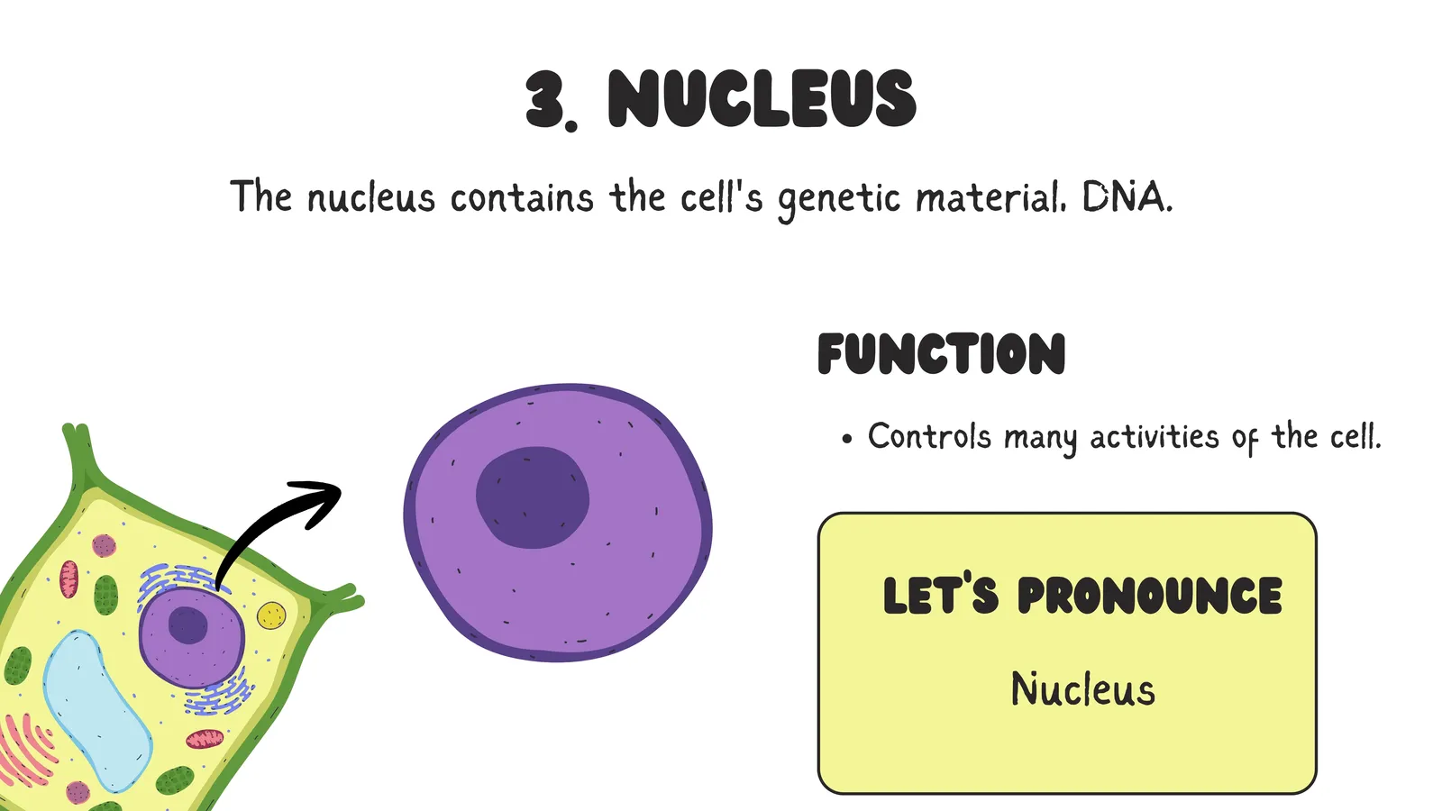
Slide: The nucleus
The control centre
1/1
Mitochondria: The Powerhouse of the Cell
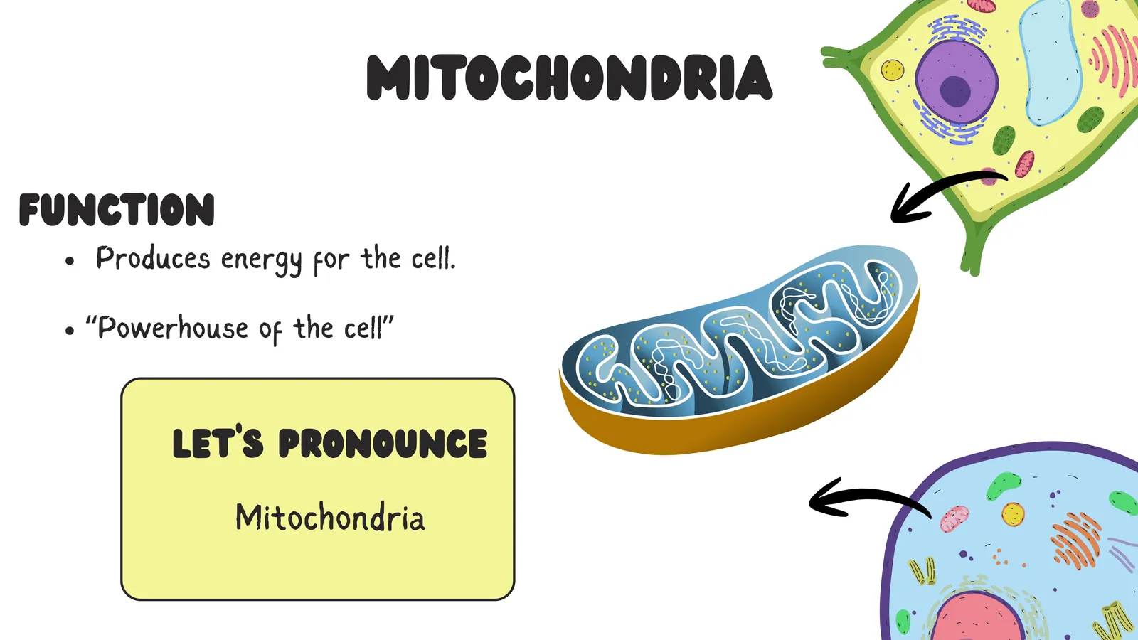
Mitochondria are rod shaped or sausage shaped organelles found in nearly all eukaryotic cells. The singular form is mitochondrion, and getting that right is worth a mark in many examinations.
Structure of Mitochondria
A mitochondrion has two membranes. The outer membrane is smooth and forms the boundary. The inner membrane is deeply folded into finger like projections called cristae, and the space enclosed by it is filled with a fluid called the matrix.
Those folds are not decoration. The enzymes of respiration are embedded in the inner membrane, so folding it increases the working surface area enormously without increasing the size of the organelle. It is the same principle as villi in the intestine and alveoli in the lungs. Whenever biology needs more surface in a small space, it folds something.
Mitochondria are unusual in another way. They contain their own small circular DNA and their own ribosomes, and they can multiply by dividing independently of the cell. Many biologists believe this is because mitochondria descended from free living bacteria that were absorbed by a larger cell long ago and stayed on as permanent partners.
Function of Mitochondria
Mitochondria carry out aerobic respiration, releasing energy from glucose and storing it in a molecule called ATP. ATP is the usable energy currency of the cell, spent on muscle contraction, active transport, protein building and every other energy requiring process.
The number of mitochondria in a cell tells you how energetic that cell is. A heart muscle cell, which contracts continuously throughout your life, is packed with them. A sperm cell has a dense spiral of mitochondria in its middle piece to power the tail. A fat storage cell, which does very little work, has relatively few.
⚡
Slide: Mitochondria
The powerhouse of the cell
1/1
Ribosomes: The Protein Factories
Ribosomes are the smallest organelles and the only ones with no membrane at all. Each is made of two subunits built from RNA and protein.
Ribosomes are found in two places. Some float freely in the cytoplasm and make proteins that will be used inside the cell. Others are attached to the surface of the endoplasmic reticulum and make proteins that will be exported out of the cell.
Their function is protein synthesis. They read the coded instructions carried from the nucleus and join amino acids together in the correct order to build a protein. Because every enzyme is a protein, ribosomes are essential to the entire life of the cell.
Importantly, ribosomes are the one organelle found in both prokaryotic and eukaryotic cells, since no cell can survive without making proteins. Prokaryotic ribosomes are smaller, a difference some antibiotics exploit by attacking bacterial ribosomes while leaving human ones alone.
Endoplasmic Reticulum: The Transport Network
The endoplasmic reticulum, usually shortened to ER, is a vast network of flattened sacs and tubes extending through the cytoplasm and connected to the nuclear envelope. It comes in two forms.
Rough Endoplasmic Reticulum
The rough ER is studded with ribosomes on its outer surface, which gives it a bumpy appearance under an electron microscope and explains its name.
Its function is to collect, fold and transport the proteins made by those attached ribosomes. Proteins destined for export are pushed into the interior of the rough ER, folded into their correct shape, and then packaged into small bubbles called vesicles that bud off and travel onwards.
Cells that produce large amounts of protein for export, such as pancreatic cells making digestive enzymes or white blood cells making antibodies, contain a great deal of rough ER.
Smooth Endoplasmic Reticulum
The smooth ER has no ribosomes attached. It handles lipids rather than proteins, making fats, oils, phospholipids and steroid hormones. It also stores calcium ions and, in liver cells, plays a major role in detoxifying alcohol, drugs and other harmful substances.
This is why liver cells and the cells of hormone producing glands such as the ovaries and testes contain unusually large amounts of smooth ER.
Golgi Apparatus: The Packaging and Dispatch Department
The Golgi apparatus, also called the Golgi body or Golgi complex, consists of a stack of flattened membrane sacs, rather like a pile of pancakes, with small vesicles clustered around the edges. It is named after Camillo Golgi, the Italian scientist who discovered it.
Its job is to receive, modify, sort and dispatch. Vesicles arriving from the endoplasmic reticulum fuse with one side of the stack. As the contents move through, they are chemically modified, often by adding sugar groups to proteins to form glycoproteins. At the far side, finished products are packaged into new vesicles and labelled for their destination.
Some vesicles carry material to the cell membrane for release outside the cell, a process called secretion. Others become lysosomes. Others deliver enzymes to specific locations within the cell.
The best comparison is a postal sorting office. Items arrive in bulk, are checked and repackaged, are given the correct address label, and are then sent out to the right destination. Without the Golgi apparatus, the cell would produce proteins but have no reliable way to finish and deliver them.
Lysosomes: The Recycling and Demolition Crew
Lysosomes are small spherical sacs filled with powerful digestive enzymes, produced by the Golgi apparatus. They are common in animal cells and less prominent in plant cells.
Lysosomes have several roles. They digest large food particles taken into the cell so the nutrients can be used. They break down worn out organelles so their components can be recycled, which is a form of cellular housekeeping. In white blood cells, lysosomes destroy bacteria that have been engulfed.
They also carry out programmed cell destruction. When a cell is damaged, diseased or no longer needed, its lysosomes can rupture and release their enzymes, digesting the cell from within. For this reason lysosomes are sometimes called the suicide bags of the cell. This sounds alarming, but it is essential. It is how a tadpole reabsorbs its tail and how the webbing between the fingers of a human embryo disappears before birth.
Chloroplasts: The Food Producers of Plants
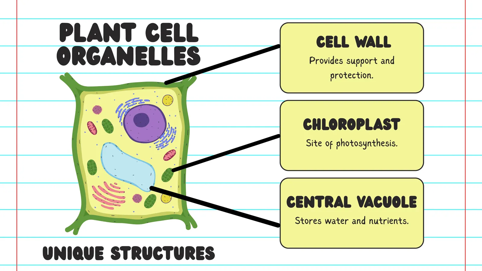
Chloroplasts are found only in plant cells and algae, specifically in the green parts exposed to light. They are lens shaped organelles surrounded by a double membrane.
Inside, the chloroplast contains stacks of disc shaped membranes called grana, and these membranes hold the green pigment chlorophyll. Around them is a fluid called stroma.
Chloroplasts carry out photosynthesis. Chlorophyll absorbs light energy, and that energy is used to combine carbon dioxide and water into glucose, releasing oxygen as a byproduct. This single process supports almost all life on Earth, because it provides both the food at the base of nearly every food chain and the oxygen that most organisms breathe.
Like mitochondria, chloroplasts contain their own DNA and ribosomes and can divide on their own, supporting the idea that they too were once independent organisms.
It is worth noting the neat relationship between the two energy organelles. Chloroplasts store energy by building glucose, while mitochondria release energy by breaking glucose down. Plant cells contain both, which is why a plant can feed itself and still respire at night when there is no light.
Vacuoles: Storage Spaces
A vacuole is a fluid filled sac surrounded by a single membrane. Its appearance differs greatly between plant and animal cells.
Plant cells usually have one very large permanent central vacuole that can take up most of the cell volume. It is filled with cell sap and enclosed by a membrane called the tonoplast. It stores water, sugars, salts and sometimes coloured pigments that give flowers their colour. Its most important structural role is creating turgor pressure, pushing outwards against the cell wall to keep the cell firm. When plants lose water, this pressure falls and the plant wilts.
Animal cells contain only small temporary vacuoles, used for short term storage or for moving materials.
🌿
Slide: Plant cell organelles
Structures only plant cells have
1/1
Other Structures You Should Know
Centrioles
Centrioles are small cylindrical structures found in animal cells, usually as a pair near the nucleus. During cell division they organise the spindle fibres that pull chromosomes apart, making sure each daughter cell receives the correct number. Most plant cells manage division without them.
The Cytoskeleton
The cytoskeleton is a network of protein fibres running through the cytoplasm. It maintains cell shape, anchors organelles in position, and provides tracks along which vesicles are moved. Without it, a cell would be a shapeless bag with its contents drifting randomly.
Cilia and Flagella
Cilia are short hair like projections that beat in coordinated waves. The cells lining your windpipe use them to sweep mucus and trapped dust upwards away from the lungs. Flagella are longer whip like structures used for movement, and the tail of a human sperm cell is the clearest example.
The Cell Wall
Although not strictly an organelle, the cell wall deserves mention. Made of cellulose in plants, it lies outside the cell membrane, gives the cell a fixed shape, provides support and prevents bursting. It is fully permeable, so it offers strength rather than selectivity. Fungi have cell walls made of chitin instead, and bacterial walls are made of a different material again.
Summary of Organelles and Their Functions
Nucleus stores DNA and controls all cell activities including division.
Nucleolus manufactures ribosomes inside the nucleus.
Mitochondrion carries out aerobic respiration and releases energy as ATP.
Ribosome builds proteins by joining amino acids in the correct order.
Rough endoplasmic reticulum folds and transports proteins made by attached ribosomes.
Smooth endoplasmic reticulum makes lipids and steroids and detoxifies harmful substances.
Golgi apparatus modifies, packages and dispatches proteins and forms lysosomes.
Lysosome digests food particles, worn out organelles and invading bacteria.
Chloroplast carries out photosynthesis using chlorophyll in plant cells.
Vacuole stores cell sap and maintains turgor pressure in plant cells.
Centriole organises spindle fibres during animal cell division.
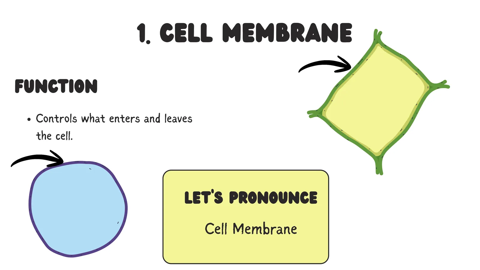
Cell membrane controls what enters and leaves the cell.
Cell wall provides rigid support in plant, fungal and bacterial cells.
🧫
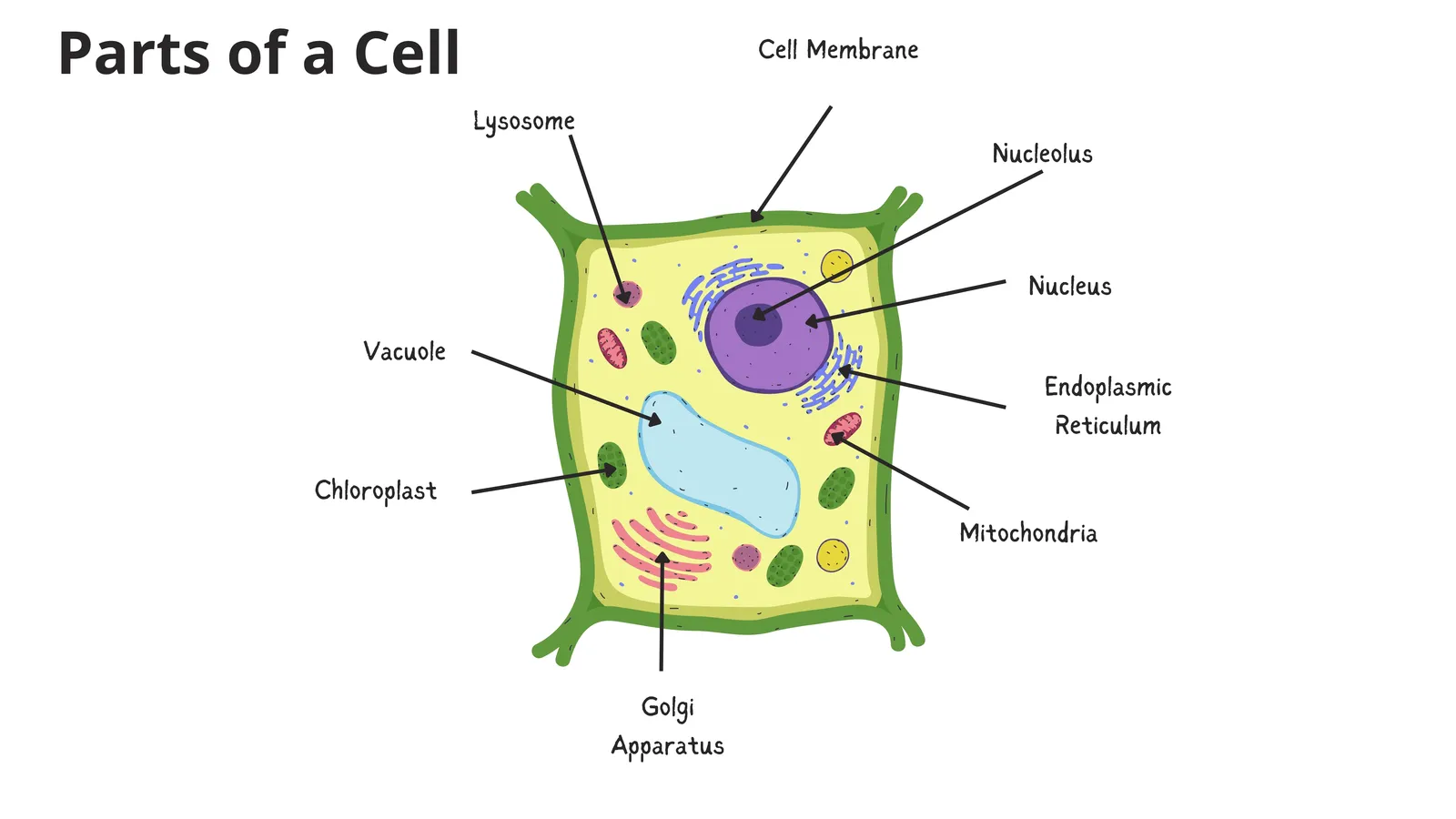
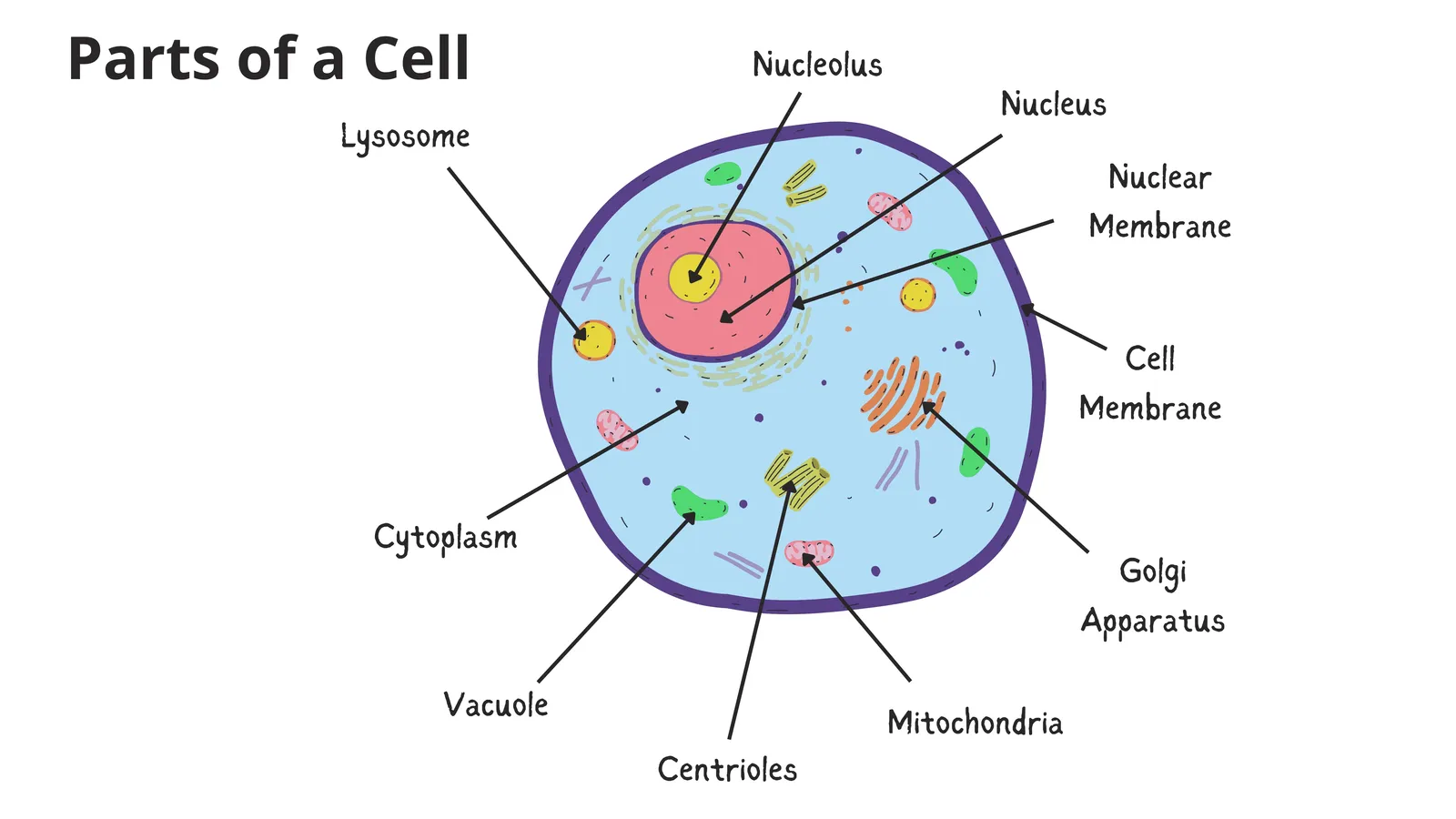
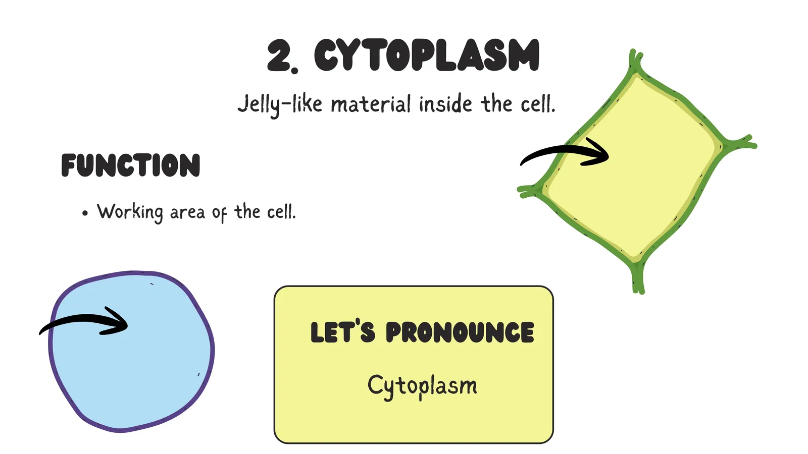
Slides: Parts of a cell
Labelled diagrams plus the cell membrane and cytoplasm
1/4
How the Organelles Work Together
Organelles do not work in isolation, and examiners love questions that ask you to trace a pathway. Follow the manufacture of a digestive enzyme in a pancreatic cell and you will see the whole system cooperating.
The instructions begin in the nucleus, where the gene for that enzyme is copied into a messenger molecule. That message leaves through a nuclear pore and reaches a ribosome attached to the rough endoplasmic reticulum. The ribosome assembles the amino acid chain, which is pushed into the rough ER and folded correctly. A vesicle buds off and carries the protein to the Golgi apparatus, which modifies it, checks it and packages it into a new vesicle with the correct destination label. That vesicle travels to the cell membrane, fuses with it, and releases the finished enzyme outside the cell. Powering every step of this journey is ATP supplied by the mitochondria.
Learn that one pathway well and you have effectively revised six organelles at once, along with the logic that connects them.
🎯
Slide: Check for understanding
Test yourself before the FAQ
1/1
Frequently Asked Questions About Cell Organelles
Why are mitochondria called the powerhouse of the cell?
Because they carry out aerobic respiration and release the energy stored in glucose, converting it into ATP, which is the form of energy the rest of the cell can actually spend.
What is the difference between rough and smooth endoplasmic reticulum?
Rough ER has ribosomes on its surface and processes proteins. Smooth ER has no ribosomes and makes lipids and steroid hormones while also detoxifying substances, especially in liver cells.
Why are lysosomes called suicide bags?
Because they contain digestive enzymes that can be released to break down the cell itself when it is damaged, diseased or no longer needed, which is a normal and necessary process during development.
Which organelles have their own DNA?
Mitochondria and chloroplasts. Both also have their own ribosomes and can divide independently, which supports the theory that they were once free living bacteria.
Which organelle is present in both plant and animal cells but does the opposite job to the chloroplast?
The mitochondrion. Chloroplasts store energy by making glucose, while mitochondria release energy by breaking glucose down.
Do plant cells have lysosomes?
Plant cells contain far fewer recognisable lysosomes, because the large central vacuole carries out much of the same digestive and storage work.
Final Thoughts
Cell organelles look intimidating at first because there are so many names to learn. The trick is to stop treating them as a vocabulary list and start treating them as a working team with a shared purpose.
Give each organelle one job in a single clear sentence. Then connect them into the protein production pathway from nucleus to ribosome to rough ER to Golgi to cell membrane. Then remember that mitochondria power the entire operation and lysosomes clean up afterwards.
Once the cell becomes a place you can picture, with departments that talk to each other, every question about organelles turns into a question you can reason through rather than one you have to recall word for word. That shift from memorising to understanding is the single most valuable study skill in biology, and this topic is the perfect place to practise it.
🗂️ Revision Flashcards
Tap a card to reveal the answer.
🎯 Quick Quiz
8 questions. Pick an answer to check it straight away.
1Which organelle has no membrane at all?
Ribosomes are the smallest organelles and the only ones with no membrane.
2Why is the inner membrane of a mitochondrion folded into cristae?
Folding increases the working surface area without increasing the size of the organelle.
3What is the job of the nucleolus?
The nucleolus makes ribosomes, which are exported into the cytoplasm.
4Which two organelles contain their own DNA and ribosomes?
Both can divide independently, supporting the idea they were once free living bacteria.
5What does smooth endoplasmic reticulum do?
Smooth ER has no ribosomes and handles lipids rather than proteins.
6Which cell would contain the most mitochondria?
Heart muscle contracts continuously, so it needs lots of energy; fat storage cells do very little work.
7What is the correct pathway for making and exporting a digestive enzyme?
Instructions leave the nucleus, the protein is built on the rough ER, finished in the Golgi and released at the membrane.
8Why are lysosomes called suicide bags?
When a cell is damaged, diseased or no longer needed, lysosomes rupture and digest it.